


-
摘要: 单颗粒示踪(Single particle tracking,SPT)技术是应用显微镜系统对细胞内单个特定荧光或散射颗粒的定位和追踪。由于SPT能够实时监控活细胞内复杂、高度动态的组织结构的变化并提供结构—功能间的动力学关系,因此在细胞生物学上有重要的应用。本文总结了SPT的机理以及在细胞上的应用,首先介绍了SPT的动力学原理,包括单颗粒定位,轨道重建以及轨道分析,然后总结了SPT技术现阶段重点发展的光学材料及仪器,最后阐述了SPT在细胞膜、细胞内信号通路、分子转运机制、遗传信息表达以及病毒感染机制的应用。此外,本文还对SPT技术未来的发展进行了展望。Abstract: Single particle tracking(SPT) technique locates and tracks individual fluorescent or scattering particles within a cell with the help of microscope system. Based on the ability of real-time monitoring of the complex and highly dynamic changes in tissue structure within living cells and the ability to provide dynamic relationships between structure and function, SPT has important applications in cell biology. In this review, the mechanism of SPT and its application on cells are summarized. Firstly, the dynamics of SPT are introduced, including single particle localization, trajectory reconstruction and analysis. Then the optical materials and instruments that SPT technology focuses on at the present stage are described. Finally, the application of SPT in cell membrane, intracellular signaling pathway, molecular transport mechanism, genetic information expression, and viral infection mechanism are proposed. In addition, the advance of SPT technology are prospected in this paper.
-
Key words:
- SPT technology /
- cell /
- dynamics /
- optical materials and instruments
-

图 1 SPT轨迹重建示意图[27]。(a)借助PSF确定颗粒的位置; (b)只和邻近的点进行连接; (c)按照时间顺序连接形成一个轨迹; (d)对轨迹进行数据分析,提取出动力学信息
Figure 1. Schematic representation of SPT[27]. (a)Particles are localized by finding the central locations of their point spread functions; (b)localizations belonging to the same particle at different times are connected using algorithms such as nearest neighbor; (c)chronological series of linked localizations form a time series trajectory; (d)dynamic information is extracted based on a statistical analysis of the trajectories


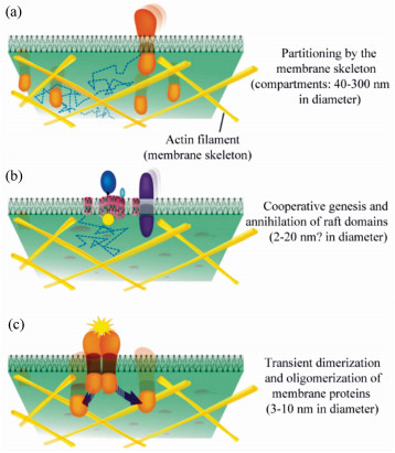
图 4 细胞膜的分层结构[17]。(a)膜隔室层,由肌动蛋白骨架分隔整个细胞膜形成,并且跨膜蛋白锚定在肌动蛋白骨架上; (b)“筏域”, 富含胆固醇,尺寸受到膜隔室的限制; (c)由膜相关蛋白以及整合蛋白的低聚物组成,存在时间非常短
Figure 4. Three-tiered hierarchical structure of mesoscale domains in the plasma membrane[17]. (a)Membrane compartments which stem from the partitioning of the entire plasma membrane by the membrane associated actin-based membrane skeleton(fence) and TM proteins anchored to the membrane skeleton fence(pickets); (b)cholesterol-containing raft domains, with sizes limited by the membrane compartments; (c)dimers and greater oligomers of membane associated and integral membrane proteins, which might exist only transiently

图 5 单个QD-驱动蛋白在活细胞内的运动[20]。(a)Hela细胞的明场图像;(b)将连续的600帧图像叠加得到的最终图像,线性轨迹表示单个QD-驱动蛋白做定向运动(实心箭头),空心箭头表示的是其他一些QD-驱动蛋白做随机运动
Figure 5. Single QD-kinesin motions in a living cell[20]. (a)Bright-field image of a HeLa cell; (b)image obtained by superimposing the 600 consecutives frames in the image sequence. The linear traject-ories are indicative of directed motions of individual QD-kinesin. Examples are marked by the full arrows. The trajectories of diffusing QD-Ks(marked by empty arrowheads) have a random shape in the superimposed image
-
[1] LIPPINCOTT-SCHWARTZ J, SNAPP E, KENWORTHY A. Studying protein dynamics in living cells[J]. Nat. Rev. Mol. Cell Bio., 2001, 2(6):444-456. doi: 10.1038/35073068 [2] REITS E A J, NEEFJES J J. From fixed to FRAP:measuring protein mobility and activity in living cells[J]. Nat. Cell Biol., 20013(6):E145-E147. doi: 10.1038/35078615 [3] DOMINGUEZ-MEDINA S, CHEN S, BLANKENBURG J, et al.. Measuring the hydrodynamic size of nanoparticles using fluctuation correlation spectroscopy[J]. Annual Review of Physical Chemistry, 2016, 67:489-514. doi: 10.1146/annurev-physchem-040214-121510 [4] YU TA, MARTINEZ MM, PAPPAS D. Fluorescence correlation spectroscopy:a review of biochemical and microfluidic applications[J]. Applied Spectroscopy, 2011, 65(4):115a-124a. doi: 10.1366/10-06224 [5] SPRAGUE B L, PEGO R L, STAVREVA DA, et al.. Analysis of binding reactions by fluorescence recovery after photobleaching[J]. Biophysical Journal, 2004, 86(6):3473-3495. doi: 10.1529/biophysj.103.026765 [6] ELSON E L. Fluorescence correlation spectroscopy:past, present, future[J]. Biophysical Journal, 2011, 101(12):2855-2870. doi: 10.1016/j.bpj.2011.11.012 [7] HAUSTEIN E, SCHWILLE P. Fluorescence correlation spectroscopy:novel variations of an established technique[J]. Annual Review of Biophysics and Biomolecular Structure, 2007, 36:151-169. doi: 10.1146/annurev.biophys.36.040306.132612 [8] GEERTS H, DE BRABANDER M, NUYDENS R, et al.. Nanovid tracking:a new automatic method for the study of mobility in living cells based on colloidal gold and video microscopy[J]. Biophysical Journal, 1987, 52(5):775-782. doi: 10.1016/S0006-3495(87)83271-X [9] DE BRABANDER M, NUYDENS R, GEERTS H, et al.. Dynamic behavior of the transferrin receptor followed in living epidermoid carcinoma(A431) cells with nanovid microscopy[J]. Cell Motility and the Cytoskeleton, 1988, 9(1):30-47. doi: 10.1002/(ISSN)1097-0169 [10] DE BRABANDER M, NUYDENS R, ISHIHARA A, et al.. Lateral diffusion and retrograde movements of individual cell surface components on single motile cells observed with Nanovid microscopy[J]. The Journal of Cell Biology, 1991, 112(1):111-124. doi: 10.1083/jcb.112.1.111 [11] BETZIG E, CHICHESTER R J. Single molecules observed by near-field scanning optical microscopy[J]. Science, 1993, 262(5138):1422-1425. doi: 10.1126/science.262.5138.1422 [12] SAKO Y, MINOGHCHI S, YANAGIDA T. Single-molecule imaging of EGFR signalling on the surface of living cells[J]. Nature Cell Biology, 2000, 2(3):168-172. doi: 10.1038/35004044 [13] ⅡNO R, KOYAMA I, KUSUMI A. Single molecule imaging of green fluorescent proteins in living cells:E-cadherin forms oligomers on the free cell surface[J]. Biophysical Journal, 2001, 80(6):2667-2677. http://www.cell.com/biophysj/pdf/S0006-3495(01)76236-4.pdf [14] SCHMIDT T, SCHUTZ GJ, BAUMGARTNER W, et al.. Imaging of single molecule diffusion[J]. Proceedings of the National Academy of Sciences of the United States of America, 1996, 93(7):2926-2929. doi: 10.1073/pnas.93.7.2926 [15] SCHUTZ G J, KADA G, PASTUSHENKO V P, et al.. Properties of lipid microdomains in a muscle cell membrane visualized by single molecule microscopy[J]. The EMBO Journal, 2000, 19(5):892-901. doi: 10.1093/emboj/19.5.892 [16] DAHAN M, LEVI S, LUCCARDINI C, et al.. Diffusion dynamics of glycine receptors revealed by single-quantum dot tracking[J]. Science, 2003, 302(5644):442-445. doi: 10.1126/science.1088525 [17] KUSUMI A, SUZUKI K G, KASAI R S, et al.. Hierarchical mesoscale domain organization of the plasma membrane[J]. Trends in Biochemical Sciences, 2011, 36(11):604-615. doi: 10.1016/j.tibs.2011.08.001 [18] WARSHAW D M, KENNEDY G G, WORK S S, et al.. Differential labeling of myosin V heads with quantum dots allows direct visualization of hand-over-hand processivity[J]. Biophysical Journal, 2005, 88(5):L30-32. doi: 10.1529/biophysj.105.061903 [19] NAN X, SIMS P A, CHEN P, et al.. Observation of individual microtubule motor steps in living cells with endocytosed quantum dots[J]. The Journal of Physical Chemistry B, 2005, 109(51):24220-24224. doi: 10.1021/jp056360w [20] COURTY S, LUCCARDINI C, BELLAICHE Y, et al.. Tracking individual kinesin motors in living cells using single quantum-dot imaging[J]. Nano Letters, 2006, 6(7):1491-1495. doi: 10.1021/nl060921t [21] BIERMANN B, SOKOLL S, KLUEVA J, et al.. Imaging of molecular surface dynamics in brain slices using single-particle tracking[J]. Nature Communications, 2014, 5. http://cn.bing.com/academic/profile?id=c801e105fb5de70e3ee952400841fee7&encoded=0&v=paper_preview&mkt=zh-cn [22] NAM S H, BAE Y M, PARK Y I, et al.. Long-term real-time tracking of lanthanide ion doped upconverting nanoparticles in living cells[J]. Angewandte Chemie, 2011, 50(27):6093-6097. doi: 10.1002/anie.v50.27 [23] KURAL C, KIM H, SYED S, et al.. Kinesin and dynein move a peroxisome in vivo:a tug-of-war or coordinated movement?[J]. Science, 2005, 308(5727):1469-1472. doi: 10.1126/science.1108408 [24] YILDIZ A, FORKEY J N, MCKINNEY S A, et al.. Myosin V walks hand-over-hand:single fluorophore imaging with 1.5-nm localization[J]. Science, 2003, 300(5628):2061-2065. doi: 10.1126/science.1084398 [25] CHEN K, GU Y, SUN W, et al.. Characteristic rotational behaviors of rod-shaped cargo revealed by automated five-dimensional single particle tracking[J]. Nature Communications, 2017, 8(1):887. doi: 10.1038/s41467-017-01001-9 [26] CHEEZUM M K, WALKER W F, GUILFORD W H. Quantitative comparison of algorithms for tracking single fluorescent particles[J]. Biophysical Journal, 2001, 81(4):2378-2388. doi: 10.1016/S0006-3495(01)75884-5 [27] SHEN H, TAUZIN L J, BAIYASI R, et al.. Single particle tracking:from theory to biophysical applications[J]. Chemical Reviews, 2017, 117(11):7331-7376. doi: 10.1021/acs.chemrev.6b00815 [28] MANZO C, GARCIA-PARAJO M F. A review of progress in single particle tracking:from methods to biophysical insights[J]. Reports on Progress in Physics. Physical Society, 2015, 78(12):124601. doi: 10.1088/0034-4885/78/12/124601 [29] SERGE A, BERTAUX N, RIGNEAULT H, et al.. Dynamic multiple-target tracing to probe spatiotemporal cartography of cell membranes[J]. Nature Methods, 2008, 5(8):687-694. doi: 10.1038/nmeth.1233 [30] SAXTON M J, JACOBSON K. Single-particle tracking:applications to membrane dynamics[J]. Annual Review of Biophysics and Biomolecular Structure, 1997, 26:373-399. doi: 10.1146/annurev.biophys.26.1.373 [31] DEAN K M, PALMER A E. Advances in fluorescence labeling strategies for dynamic cellular imaging[J]. Nature Chemical Biology, 2014, 10(7):512-523. doi: 10.1038/nchembio.1556 [32] FERNANDEZ-SUAREZ M, TING A Y. Fluorescent probes for super-resolution imaging in living cells[J]. Nature Reviews. Molecular Cell Biology, 2008, 9(12):929-943. doi: 10.1038/nrm2531 [33] SHEETZ M P, TURNEY S, QIAN H, et al.. Nanometre-level analysis demonstrates that lipid flow does not drive membrane glycoprotein movements[J]. Nature, 1989, 340(6231):284-288. doi: 10.1038/340284a0 [34] GARCIA-PARAJO M F, SEGERS-NOLTEN G M, VEERMAN J A, et al.. Real-time light-driven dynamics of the fluorescence emission in single green fluorescent protein molecules[J]. Proceedings of the National Academy of Sciences of the United States of America, 2000, 97(13):7237-7242. doi: 10.1073/pnas.97.13.7237 [35] BRUCHEZ M J R, MORONNE M, GIN P, et al. Semiconductor nanocrystals as fluorescent biological labels[J]. Science, 1998, 281(5385):2013-2016. doi: 10.1126/science.281.5385.2013 [36] YAO J, LARSON D R, VISHWASRAO H D, et al.. Blinking and nonradiant dark fraction of water-soluble quantum dots in aqueous solution[J]. Proceedings of the National Academy of Sciences of the United States of America, 2005, 102(40):14284-14289. doi: 10.1073/pnas.0506523102 [37] DERFUS A M, CHAN W C W, BHATIA S N. Probing the cytotoxicity of semiconductor quantum dots[J]. Nano Letters, 2004, 4(1):11-18. doi: 10.1021/nl0347334 [38] MIAO P, HAN K, TANG Y G, et al.. Recent advances in carbon nanodots:synthesis, properties and biomedical applications[J]. Nanoscale, 2015, 7(5):1586-1595. doi: 10.1039/C4NR05712K [39] MIAO P, TANG Y G, HANA K, et al.. Facile synthesis of carbon nanodots from ethanol and their application in ferric(Ⅲ) ion assay[J]. J. Mater. Chem. A, 2015, 3(29):15068-15073. doi: 10.1039/C5TA03278D [40] CHANG Y R, LEE H Y, CHEN K, et al.. Mass production and dynamic imaging of fluorescent nanodiamonds[J]. Nature Nanotechnology, 2008, 3(5):284-288. doi: 10.1038/nnano.2008.99 [41] FU C C, LEE H Y, CHEN K, et al.. Characterization and application of single fluorescent nanodiamonds as cellular biomarkers[J]. Proceedings of the National Academy of Sciences of the United States of America, 2007, 104(3):727-732. doi: 10.1073/pnas.0605409104 [42] JIN H, HELLER D A, STRANO M S. Single-particle tracking of endocytosis and exocytosis of single-walled carbon nanotubes in NIH-3T3 cells[J]. Nano Letters, 2008, 8(6):1577-1585. doi: 10.1021/nl072969s [43] JIN H, HELLER D A, SHARMA R, et al. Size-dependent cellular uptake and expulsion of single-walled carbon nanotubes:single particle tracking and a generic uptake model for nanoparticles[J]. ACS Nano, 2009, 3(1):149-158. doi: 10.1021/nn800532m [44] WU S, HAN G, MILLIRON D J, et al.. Non-blinking and photostable upconverted luminescence from single lanthanide-doped nanocrystals[J]. Proceedings of the National Academy of Sciences of the United States of America, 2009, 106(27):10917-10921. doi: 10.1073/pnas.0904792106 [45] BAE Y M, PARK Y I, NAM S H, et al.. Endocytosis, intracellular transport, and exocytosis of lanthanide-doped upconverting nanoparticles in single living cells[J]. Biomaterials, 2012, 33(35):9080-9086. doi: 10.1016/j.biomaterials.2012.08.039 [46] WANG F, HAN Y, LIM C S, et al.. Simultaneous phase and size control of upconversion nanocrystals through lanthanide doping[J]. Nature, 2010, 463(7284):1061-1065. doi: 10.1038/nature08777 [47] LIU X J, TU Y, GAI H W. Imaging of single molecules by wide-field optical microscopy[J]. Prog. Chem., 2013, 25(2-3):370-379. doi: 10.7536/PC120842 [48] MICHALET X, COLYER R A, SCALIA G, et al.. Development of new photon-counting detectors for single-molecule fluorescence microscopy[J]. Philos. T. R. Soc. B, 2013, 368(1611) http://cn.bing.com/academic/profile?id=aae4dbdd448b3ebf29f19710702ad390&encoded=0&v=paper_preview&mkt=zh-cn [49] WIESER S, SCHUTZ G J. Tracking single molecules in the live cell plasma membrane-Do's and Don't's[J]. Methods, 2008, 46(2):131-140. doi: 10.1016/j.ymeth.2008.06.010 [50] MICHALET X, SIEGMUND O H W, VALLERGA J V, et al.. Detectors for single-molecule fluorescence imaging and spectroscopy[J]. J. Mod. Optic, 2007, 54(2-3):239-281. doi: 10.1080/09500340600769067 [51] AXELROD D. Total internal reflection fluorescence microscopy in cell biology[J]. Traffic, 2001, 2(11):764-774. doi: 10.1034/j.1600-0854.2001.21104.x [52] TOKUNAGA M, IMAMOTO N, SAKATA-SOGAWA K. Highly inclined thin illumination enables clear single-molecule imaging in cells[J]. Nature Methods, 2008, 5(2):159-161. doi: 10.1038/nmeth1171 [53] STENDER A S, MARCHUK K, LIU C, et al.. Single cell optical imaging and spectroscopy[J]. Chemical Reviews, 2013, 113(4):2469-2527. doi: 10.1021/cr300336e [54] ARHEL N, GENOVESIO A, KIM K A, et al.. Quantitative four-dimensional tracking of cytoplasmic and nuclear HIV-1 complexes[J]. Nature Methods, 2006, 3(10):817-824. doi: 10.1038/nmeth928 [55] LANGE S, KATAYAMA Y, SCHMID M, et al.. Simultaneous transport of different localized mRNA species revealed by live-cell imaging[J]. Traffic, 2008, 9(8):1256-1267. doi: 10.1111/tra.2008.9.issue-8 [56] MANLEY S, GILLETTE J M, PATTERSON G H, et al.. High-density mapping of single-molecule trajectories with photoactivated localization microscopy[J]. Nature Methods, 2008, 5(2):155-157. doi: 10.1038/nmeth.1176 [57] PASZEK M J, DUFORT C C, ROSSIER O, et al.. The cancer glycocalyx mechanically primes integrin-mediated growth and survival[J]. Nature, 2014, 511(7509):319-325. doi: 10.1038/nature13535 [58] ROSSIER O, OCTEAU V, SIBARITA J B, et al.. Integrins beta1 and beta3 exhibit distinct dynamic nanoscale organizations inside focal adhesions[J]. Nature Cell Biology, 2012, 14(10):1057-1067. doi: 10.1038/ncb2588 [59] GIANNONE G, HOSY E, LEVET F, et al.. Dynamic superresolution imaging of endogenous proteins on living cells at ultra-high density[J]. Biophysical Journal, 2010, 99(4):1303-1310. doi: 10.1016/j.bpj.2010.06.005 [60] GARCIA-PARAJO M F, CAMBI A, TORRENO-PINA J A, et al.. Nanoclustering as a dominant feature of plasma membrane organization[J]. Journal of Cell Science, 2014, 127(Pt 23):4995-5005. http://cn.bing.com/academic/profile?id=9eda786cb0e24492fe6eb11becba9b10&encoded=0&v=paper_preview&mkt=zh-cn [61] LINGWOOD D, SIMONS K. Lipid rafts as a membrane-organizing principle[J]. Science, 2010, 327(5961):46-50. doi: 10.1126/science.1174621 [62] KUSUMI A, TSUNOYAMA T A, HIROSAWA K M, et al.. Tracking single molecules at work in living cells[J]. Nature Chemical Biology, 2014, 10(7):524-532. doi: 10.1038/nchembio.1558 [63] KUSUMI A, NAKADA C, RITCHIE K, et al.. Paradigm shift of the plasma membrane concept from the two-dimensional continuum fluid to the partitioned fluid:high-speed single-molecule tracking of membrane molecules[J]. Annual Review of Biophysics and Biomolecular Structure, 2005, 34:351-378. doi: 10.1146/annurev.biophys.34.040204.144637 [64] SAKO Y, KUSUMI A. Barriers for lateral diffusion of transferrin receptor in the plasma membrane as characterized by receptor dragging by laser tweezers:fence versus tether[J]. The Journal of Cell Biology, 1995, 129(6):1559-1574. doi: 10.1083/jcb.129.6.1559 [65] FUJIWARA T, RITCHIE K, MURAKOSHI H, et al.. Phospholipids undergo hop diffusion in compartmentalized cell membrane[J]. The Journal of Cell Biology, 2002, 157(6):1071-1081. doi: 10.1083/jcb.200202050 [66] SUZUKI K G, KASAI R S, HIROSAWA K M, et al.. Transient GPI-anchored protein homodimers are units for raft organization and function[J]. Nature Chemical Biology, 2012, 8(9):774-783. doi: 10.1038/nchembio.1028 [67] ANDREWS N L, LIDKE K A, PFEIFFER J R, et al.. Actin restricts FcepsilonRI diffusion and facilitates antigen-induced receptor immobilization[J]. Nature Cell Biology, 2008, 10(8):955-963. doi: 10.1038/ncb1755 [68] OWEN D M, WILLIAMSON D, RENTERO C, et al.. Quantitative microscopy:protein dynamics and membrane organisation[J]. Traffic, 2009, 10(8):962-971. doi: 10.1111/tra.2009.10.issue-8 [69] TREANOR B, DEPOIL D, GONZALEZ-GRANJA A, et al.. The membrane skeleton controls diffusion dynamics and signaling through the B cell receptor[J]. Immunity, 2010, 32(2):187-199. doi: 10.1016/j.immuni.2009.12.005 [70] MURASE K, FUJIWARA T, UMEMURA Y, et al.. Ultrafine membrane compartments for molecular diffusion as revealed by single molecule techniques[J]. Biophysical Journal, 2004, 86(6):4075-4093. doi: 10.1529/biophysj.103.035717 [71] WIESER S, MOERTELMAIER M, FUERTBAUER E, et al.. (Un)confined diffusion of CD59 in the plasma membrane determined by high-resolution single molecule microscopy[J]. Biophysical Journal, 2007, 92(10):3719-3728. doi: 10.1529/biophysj.106.095398 [72] WEGNER K D, HILDEBRANDT N. Quantum dots:bright and versatile in vitro and in vivo fluorescence imaging biosensors[J]. Chem. Soc. Rev., 2015, 44(14):4792-4834. doi: 10.1039/C4CS00532E [73] LEDUC C, SI S, GAUTIER J, et al.. A highly specific gold nanoprobe for live-cell single-molecule imaging[J]. Nano Letters, 2013, 13(4):1489-1494. doi: 10.1021/nl304561g [74] TAURAN Y, BRIOUDE A, COLEMAN A W, et al.. Molecular recognition by gold, silver and copper nanoparticles[J]. World Journal of Biological Chemistry, 2013, 4(3):35-63. doi: 10.4331/wjbc.v4.i3.35 [75] LIDKE DS, LIDKE KA, RIEGER B, et al.. Reaching out for signals:filopodia sense EGF and respond by directed retrograde transport of activated receptors[J]. The Journal of Cell Biology, 2005, 170(4):619-626. doi: 10.1083/jcb.200503140 [76] RAJAN S S, LIU H Y, VU T Q. Ligand-bound quantum dot probes for studying the molecular scale dynamics of receptor endocytic trafficking in live cells[J]. ACS Nano, 2008, 2(6):1153-1166. doi: 10.1021/nn700399e [77] BHATIA D, ARUMUGAM S, NASILOWSKI M, et al.. Quantum dot-loaded monofunctionalized DNA icosahedra for single-particle tracking of endocytic pathways[J]. Nature Nanotechnology, 2016, 11(12):1112-1119. doi: 10.1038/nnano.2016.150 [78] PIEROBON P, ACHOURI S, COURTY S, et al.. Velocity, processivity, and individual steps of single myosin V molecules in live cells[J]. Biophysical Journal, 2009, 96(10):4268-4275. doi: 10.1016/j.bpj.2009.02.045 [79] FAKHRI N, WESSEL A D, WILLMS C, et al.. High-resolution mapping of intracellular fluctuations using carbon nanotubes[J]. Science, 2014, 344(6187):1031-1035. doi: 10.1126/science.1250170 [80] BALINT S, VERDENY V I, SANDOVAL A A, et al. Correlative live-cell and superresolution microscopy reveals cargo transport dynamics at microtubule intersections[J]. Proceedings of the National Academy of Sciences of the United States of America, 2013, 110(9):3375-3380. doi: 10.1073/pnas.1219206110 [81] APPELHANS T, RICHTER C P, WILKENS V, et al.. Nanoscale organization of mitochondrial microcompartments revealed by combining tracking and localization microscopy[J]. Nano Letters, 2012, 12(2):610-616. doi: 10.1021/nl203343a [82] YOO J, KAMBARA T, GONDA K, et al.. Intracellular imaging of targeted proteins labeled with quantum dots[J]. Experimental Cell Research, 2008, 314(19):3563-3569. doi: 10.1016/j.yexcr.2008.09.014 [83] VALM A M, COHEN S, LEGANT W R, et al.. Applying systems-level spectral imaging and analysis to reveal the organelle interactome[J]. Nature, 2017, 546(7656):162-167. doi: 10.1038/nature22369 [84] MISTELI T. Beyond the sequence:cellular organization of genome function[J]. Cell, 2007, 128(4):787-800. doi: 10.1016/j.cell.2007.01.028 [85] HANDWERGER K E, GALL J G. Subnuclear organelles:new insights into form and function[J]. Trends in Cell Biology, 2006, 16(1):19-26. doi: 10.1016/j.tcb.2005.11.005 [86] VIVANTE A, BROZGOL E, BRONSHTEIN I, et al.. Genome organization in the nucleus:from dynamic measurements to a functional model[J]. Methods, 2017, 123:128-137. doi: 10.1016/j.ymeth.2017.01.008 [87] LEVI V, RUAN Q, PLUTZ M, et al.. Chromatin dynamics in interphase cells revealed by tracking in a two-photon excitation microscope[J]. Biophysical Journal, 2005, 89(6):4275-4285. doi: 10.1529/biophysj.105.066670 [88] GEBHARDT J C, SUTER DM, ROY R, et al. Single-molecule imaging of transcription factor binding to DNA in live mammalian cells[J]. Nature Methods, 2013, 10(5):421-426. doi: 10.1038/nmeth.2411 [89] LOWE A R, SIEGEL J J, KALAB P, et al.. Selectivity mechanism of the nuclear pore complex characterized by single cargo tracking[J]. Nature, 2010, 467(7315):600-603. doi: 10.1038/nature09285 [90] LIU H B, LIU Y, LIU S L, et al.. Clathrin-mediated endocytosis in living host cells visualized through quantum dot labeling of infectious hematopoietic necrosis virus[J]. J. Virol., 2011, 85(13):6252-6262. doi: 10.1128/JVI.00109-11 [91] WEN L, LIN Y, ZHENG ZH, et al.. Labeling the nucleocapsid of enveloped baculovirus with quantum dots for single-virus tracking[J]. Biomaterials, 2014, 35(7):2295-2301. doi: 10.1016/j.biomaterials.2013.11.069 [92] LV C, LIN Y, LIU A A, et al.. Labeling viral envelope lipids with quantum dots by harnessing the biotinylated lipid-self-inserted cellular membrane[J]. Biomaterials, 2016, 106:69-77. doi: 10.1016/j.biomaterials.2016.08.013 [93] JOO K I, FANG Y, LIU Y, et al.. Enhanced real-time monitoring of adeno-associated virus trafficking by virus-quantum dot conjugates[J]. ACS Nano, 2011, 5(5):3523-3535. doi: 10.1021/nn102651p [94] LIU S L, ZHANG Z L, TIAN Z Q, et al.. Effectively and efficiently dissecting the infection of influenza virus by quantum-dot-based single-particle tracking[J]. ACS Nano, 2012, 6(1):141-150. doi: 10.1021/nn2031353 [95] LI Q, LI W, YIN W, et al.. Single-particle tracking of human immunodeficiency virus type 1 productive entry into human primary macrophages[J]. ACS Nano, 2017, 11(4):3890-3903. doi: 10.1021/acsnano.7b00275 [96] YU J, ZHANG X J, HAO X J, et al.. Near-infrared fluorescence imaging using organic dye nanoparticles[J]. Biomaterials, 2014, 35(10):3356-3364. doi: 10.1016/j.biomaterials.2014.01.004 [97] NI M, ZHUO S, SO P T, et al.. Fluorescent probes for nanoscopy:four categories and multiple possibilities[J]. Journal of Biophotonics, 2017, 10(1):11-23. doi: 10.1002/jbio.201600042 [98] NOLLE J M, PRIMPKE S, MULLEN K, et al.. Diffusion of single molecular and macromolecular probes during the free radical bulk polymerization of MMA-towards a better understanding of the Trommsdorff effect on a molecular level[J]. Polym. Chem.-Uk, 2016, 7(24):4100-4105. doi: 10.1039/C6PY00590J [99] HIGGINS D A, PARK S C, TRAN-BA K H, et al.. Single-molecule investigations of morphology and mass transport dynamics in nanostructured materials[J]. Annu. Rev. Anal. Chem., 2015, 8:193-216. doi: 10.1146/annurev-anchem-071114-040153 -

 下载:
下载:


 点击查看大图

计量
- 文章访问数: 3784
- HTML全文浏览量: 1298
- PDF下载量: 498
- 被引次数: 0
