


Application in nucleic acid functionalized nanoprobe in cellular fluorescence imaging
-
摘要: 核酸是携带遗传信息的物质,既存在于自然界中也能够通过成熟技术人工合成。通过体外筛选技术还可以筛选出具有特殊功能的核酸序列,例如核酸适体和脱氧核酶。核酸通过沃森-克里克碱基互补配对原则进行杂交,具有很强的专一性。无论是通过序列设计还是体外筛选,核酸探针在生物标志物的分析与成像应用方面都发挥着重要作用。纳米材料辅助构建核酸功能化纳米探针,可以保护负载的核酸探针不被核酸酶降解,并且无需转染试剂就能进入细胞,在细胞荧光成像应用上具有很大优势。为解决细胞内有些生物标志物含量低、难于检测的问题,目前已构建多种适用于细胞水平的成像信号放大方法来实现对低丰度生物标志物的高灵敏成像。本文主要综述了核酸功能化纳米探针在细胞荧光成像中的应用进展,包括反义寡核苷酸功能化纳米探针、核酸适体功能化纳米探针、脱氧核酶功能化纳米探针等,同时介绍了他们在成像信号放大中的应用。Abstract: Nucleic acid is a substance that carries genetic information. It exists either in nature or can be synthesized by established techniques. Nucleic acid sequences with special functions, such as aptamers and DNAzymes, can also be selected using in vitro techniques. Nucleic acids hybridize according to the principle of Watson-Crick base pairing, and have strong specificity. Whether through sequence design or in vitro screening, nucleic acid probes play an important role in the analysis and imaging applications of biomarkers. In addition, nanomaterials can be used to construct nucleic acid functionalized nanoprobes, which can protect the loaded nucleic acids from being degraded by nucleases and can enter cells without the aid of the transfection reagents. Therefore, nanomaterials have great advantages in the application of cell fluorescence imaging. In order to solve the problem of low intracellular biomarker content and thus difficult to detect, a variety of imaging signal amplification methods suitable for the cellular level have been developed to achieve highly sensitive imaging of low-abundance biomarkers. In this paper, the application of nucleic acid functionalized nanoprobes in cellular fluorescence imaging, including antisense oligonucleotide functionalized nanoprobes, aptamer functionalized nanoprobes and DNAzyme functionalized nanoprobes and that in imaging signal amplification are reviewed.
-
Key words:
- nucleic acid /
- nanoprobe /
- antisense oligonucleotide /
- aptamer /
- DNAzyme /
- fluorescence imaging
-

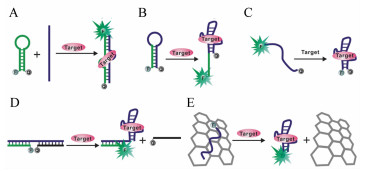
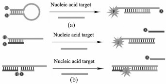
图 1 (a) 分子信标响应原理;(b)线型核酸探针响应原理
Figure 1. (a)Rresponse principle of molecular beacons; (b)Response principle of linear nucleic acid probe

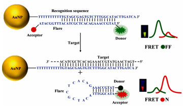
图 2 (a) 纳米金辅助构建的核酸纳米探针用于细胞内mRNA成像[18];(b)聚多巴胺辅助构建的核酸纳米探针用于成像细胞分化相关的miRNA[19];(c)多色核酸纳米探针用于活细胞内肿瘤相关mRNA成像[20];(d)核酸作为模板合成具有荧光性质的金纳米簇并用于细胞成像[21]
Figure 2. (a)AuNPs-based nucleic acid nanoprobe for application in intracellular mRNA imaging[18]; (b)polydopamine-based nucleic acid nanoprobe for the imaging of miRNAs in living human mesenchymal stem cells[19]; (c)a multicolor nanoprobe for imaging of tumor-related mRNAs in living cells[20]; (d)nucleic acid based template for the synthesis of fluorescent gold nanoclusters and their application in cell imaging[21]


图 7 (a) 金纳米棒辅助构建的核酸适体探针用于癌细胞特异性的成像与治疗[37];(b)核酸适体功能化纳米探针用于线粒体释放的细胞色素C的荧光成像[38];(c)AS1411核酸适体与反义寡核苷酸同时功能化的纳米颗粒用于癌细胞内microRNA-221荧光成像[39]
Figure 7. (a)Gold nanorods based aptamer switch probes for cancer cell specific imaging and treatment[37]; (b)fluorescence imaging of cytochrome c released from mitochondria using aptameric nanoprobe[38]; (c)fluorescence imaging of microRNA-221 in cancer cells by using nucleolin aptamer and antisense oligonucleotides functionalized nanoparticles[39]

图 8 (a) 脱氧核酶体外筛选过程;(b)脱氧核酶的结构与切割位点[42];(c)目标核酸诱导活性脱氧核酶的形成[43];(d)核酸适体与目标分子结合诱导活性脱氧核酶的形成[44]
Figure 8. (a)Schematic presentation of DNAzyme selection process; (b)structure of DNAzyme and the RNA cleavage site[42]; (c)target nucleic acid induced the formation of an active DNAzyme[43]; (d)binding of aptamer to target molecule induced the formation of an active DNAzyme[44]

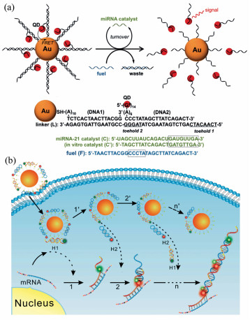
图 9 (a) 活细胞内基于脱氧核酶功能化纳米金的Zn2+和Cu2+同时荧光成像研究[45];(b)核酸适体功能化的脱氧核酶与纳米金结合用于活细胞内目标分子的放大荧光成像[46]
Figure 9. (a)Simultaneous fluorescence imaging of Zn2+ and Cu2+ in living cells based on DNAzyme modified gold nanoparticle[45]; (b)aptazyme-gold nanoparticle sensor for amplified molecular probing in living cells[46]
-
[1] TORABI S F, LU Y. Functional DNA nanomaterials for sensing and imaging in living cells[J]. Curr. Opin Biotechnol., 2014, 28:88-95. doi: 10.1016/j.copbio.2013.12.011 [2] LI J, MO L, LU C H, et al.. Functional nucleic acid-based hydrogels for bioanalytical and biomedical applications[J]. Chem. Soc. Rev., 2016, 45(5):1410-1431. doi: 10.1039/C5CS00586H [3] LIANG H, ZHANG X B, LV Y, et al.. Functional DNA-containing nanomaterials:cellular applications in biosensing, imaging, and targeted therapy[J]. Acc Chem. Res., 2014, 47(6):1891-1901. doi: 10.1021/ar500078f [4] MIAO P, TANG Y, WANG B, et al.. Near-infrared Ag2S quantum dots-based DNA logic gate platform for miRNA diagnostics[J]. Anal. Chem., 2016, 88(15):7567-7573. doi: 10.1021/acs.analchem.6b01044 [5] CHENG W, YAN W, MIAO P. TNF-α responsive DNA star trigon formation from four hairpin probes and the analytical application[J]. Sci. China. Chem., 2017, 60(3):405-409. doi: 10.1007/s11426-016-0259-4 [6] CHAKRABORTY K, VEETIL A T, JAFFREY S R, et al.. Nucleic acid-based nanodevices in biological imaging[J]. Annu. Rev. Biochem., 2016, 85(1):349-373. doi: 10.1146/annurev-biochem-060815-014244 [7] LI J, CHENG F, HUANG H, et al.. Nanomaterial-based activatable imaging probes:from design to biological applications[J]. Chem. Soc. Rev., 2015, 44(21):7855-7880. doi: 10.1039/C4CS00476K [8] LUBY B M, CHARRON D M, MACLAUGHLIN C M, et al.. Activatable fluorescence:from small molecule to nanoparticle[J]. Adv. Drug Deliv. Rev., 2016, 113:97-121. http://cn.bing.com/academic/profile?id=a7aeb37e5bac1824f6aa3ca6fbc1128f&encoded=0&v=paper_preview&mkt=zh-cn [9] HU R, ZHANG X B, KONG R M, et al.. Nucleic acid-functionalized nanomaterials for bioimaging applications[J]. J. Mater. Chem., 2011, 21(41):16323-16334. doi: 10.1039/c1jm12588e [10] ZHENG J, YANG R H, SHI M L, et al.. Rationally designed molecular beacons for bioanalytical and biomedical applications[J]. Chem. Soc. Rev., 2015, 44(10):3036-3055. doi: 10.1039/C5CS00020C [11] FARRERA C, ANDON F T, FELIU N. Carbon nanotubes as optical sensors in biomedicine[J]. ACS Nano, 2017, 11(11):10637-10643. doi: 10.1021/acsnano.7b06701 [12] ZHU X, LIU Y, LI P, et al.. Applications of graphene and its derivatives in intracellular biosensing and bioimaging[J]. Analyst, 2016, 141(15):4541-4553. doi: 10.1039/C6AN01090C [13] PENG H Y, TANG H, JIANG J H. Recent progress in gold nanoparticle-based biosensing and cellular imaging[J]. Sci. China Chem., 2016, 59(7):783-793. doi: 10.1007/s11426-016-5570-7 [14] HILDEBRANDT N, SPILLMANN C M, ALGAR W R, et al.. Energy transfer with semiconductor quantum dot bioconjugates:a versatile platform for biosensing, energy harvesting, and other developing applications[J]. Chem. Rev., 2017, 117(2):536-711. doi: 10.1021/acs.chemrev.6b00030 [15] ZHANG C, DING C, XIANG D, et al.. DNA functionalized fluorescent quantum dots for bioanalytical applications[J]. Chinese J. Chem., 2016, 34(3):317-325. doi: 10.1002/cjoc.v34.3 [16] SANTANGELO P J. Molecular beacons and related probes for intracellular RNA imaging[J]. WIREs Nanomedicine and Nanobiotechnology, 2010, 2(1):11-19. doi: 10.1002/wnan.52 [17] 杨立敏, 刘波, 李娜, 等.纳米荧光探针用于核酸分子的检测及成像研究[J].化学学报, 2017, 75:1047-1060. http://www.cnki.com.cn/Article/CJFDTotal-HXXB201711003.htmYANG L M, LIU B, LI N, et al.. Fluorescent nanoprobe for detection and imaging of nucleic acid molecules[J]. Acta Chim. Sinica, 2017, 75:1047-1060.(in Chinese) http://www.cnki.com.cn/Article/CJFDTotal-HXXB201711003.htm [18] SEFEROS D S, GILJOHANN D A, HILL H D, et al..Nano-flares:probes for transfection and mRNA detection in living cells[J]. J. Am. Chem. Soc., 2007, 129(50):15477-15479. doi: 10.1021/ja0776529 [19] CHOI C K K, LI J M, WEI K C, et al.. A gold@polydopamine core-shell nanoprobe for long-term intracellular detection of microRNAs in differentiating stem cells[J]. Methods Mol. Biol., 2017, 1570:155-164. doi: 10.1007/978-1-4939-6840-4 [20] LI N, CHANG C, PAN W, et al.. A multicolor nanoprobe for detection and imaging of tumor-related mRNAs in living cells[J]. Angew Chem. Int. Ed., 2012, 51(30):7426-7430. doi: 10.1002/anie.201203767 [21] LI J L, ZHONG X Q, CHENG F F, et al.. One-pot synthesis of aptamer-functionalized silver nanoclusters for cell-type-specific imaging[J]. Anal. Chem., 2012, 84:4140-4146. doi: 10.1021/ac3003402 [22] HE X, ZENG T, LI Z, et al.. Catalytic molecular imaging of microRNA in living cells by DNA-programmed nanoparticle disassembly[J]. Angew Chem. Int. Ed., 2016, 55(9):3073-3076. doi: 10.1002/anie.201509726 [23] YANG Y, HUANG J, YANG X, et al.. FRET nanoflares for intracellular mRNA detection:avoiding false positive signals and minimizing effects of system fluctuations[J]. J. Am. Chem. Soc., 2015, 137(26):8340-8343. doi: 10.1021/jacs.5b04007 [24] CHEN J J, TANG L J, CHU X, et al.. Enzyme-free, signal-amplified nucleic acid circuits for biosensing and bioimaging analysis[J]. Analyst, 2017, 142(7):3048-3061. http://cn.bing.com/academic/profile?id=2d3d9e3d176fe3a5cba2513c7ffb1f21&encoded=0&v=paper_preview&mkt=zh-cn [25] BI S, YUE S, ZHANG S. Hybridization chain reaction:a versatile molecular tool for biosensing, bioimaging, and biomedicine[J]. Chem. Soc. Rev., 2017, 46(14):4281-4298. doi: 10.1039/C7CS00055C [26] WU Z, LIU G Q, YANG X L, et al.. Electrostatic nucleic acid nanoassembly enables hybridization chain reaction in living cells for ultrasensitive mRNA Imaging[J]. J. Am. Chem. Soc., 2015, 137(21):6829-6836. doi: 10.1021/jacs.5b01778 [27] YANG D W, TANG Y G, MIAO P. Hybridization chain reaction directed DNA superstructures assembly for biosensing applications[J]. TrAC Trends in Analytical Chemistry, 2017, 94:1-13. doi: 10.1016/j.trac.2017.06.011 [28] MENG H M, LIU H, KUAI H, et al..Aptamer-integrated DNA nanostructures for biosensing, bioimaging and cancer therapy[J]. Chem. Soc. Rev., 2016, 45(9):2583-2602. doi: 10.1039/C5CS00645G [29] YANG D, TANG Y, GUO Z, et al.. Proximity aptasensor for protein detection based on an enzyme-free amplification strategy[J]. Mol. Bio. Syst., 2017, 13:1936-1939. http://cn.bing.com/academic/profile?id=2cc603ee3e16511018446dc02f88fb6c&encoded=0&v=paper_preview&mkt=zh-cn [30] 靳贵晓, 李娟, 杨黄浩.核酸适体的筛选及其在生物医学领域的研究进展[J].福州大学学报, 2016, 44(6):919-934. https://mall.cnki.net/qikan-SPJX201610046.htmlJIN G X, LI J, YANG H H. Research progress of aptamer screening and its application in biomedicine[J]. Journal of Fuzhou University, 2016, 44(6):919-934.(in Chinese) https://mall.cnki.net/qikan-SPJX201610046.html [31] ZHANG H, LI F, DEVER B, et al.. DNA-mediated homogeneous binding assays for nucleic acids and proteins[J]. Chem. Rev., 2013, 113(4):2812-2841. doi: 10.1021/cr300340p [32] XING H, WONG N Y, XIANG Y, et al.. DNA aptamer functionalized nanomaterials for intracellular analysis, cancer cell imaging and drug delivery[J]. Curr. Opin. Chem. Biol., 2012, 16:429-435. doi: 10.1016/j.cbpa.2012.03.016 [33] SUN H G, TAN W H, ZU Y L. Aptamers:versatile molecular recognition probes for cancer detection[J]. Analyst, 2016, 141(2):403-415. doi: 10.1039/C5AN01995H [34] LU D, HE L, ZHANG G, et al.. Aptamer-assembled nanomaterials for fluorescent sensing and imaging[J]. Nanophotonics, 2017, 6(1):109-121. http://cn.bing.com/academic/profile?id=6032d950d7888ccbaef6076faab90cde&encoded=0&v=paper_preview&mkt=zh-cn [35] DONG J T, ZHAO M P. In-vivo fluorescence imaging of adenosine 5'-triphosphate[J]. Trends in Analytical Chemistry, 2016, 80:190-203. doi: 10.1016/j.trac.2016.03.020 [36] 黄子珂, 刘超, 付强强, 等.核酸适配体荧光探针在生化分析和生物成像中的研究进展[J].应用化学, 2018, 35(1):28-39. doi: 10.11944/j.issn.1000-0518.2018.01.170363HUANG Z K, LIU CH, FU Q Q, et al.. Aptamer-based fluorescence probe for bioanalysis and bioimaging[J]. Chinese Journal of Applied Chemistry, 2018, 35(1):28-39. (in Chinese) doi: 10.11944/j.issn.1000-0518.2018.01.170363 [37] WANG J, ZHU G, YOUM, et al.. Assembly of aptamer on gold switch nanorods probes for and photosensitizer targeted photothermal and photodynamic cancer therapy[J]. ACS Nano, 2012, 6(6):5070-5077. doi: 10.1021/nn300694v [38] CHEN T T, TIAN X, LIU C L, et al.. Fluorescence activation imaging of cytochrome c released from mitochondria using aptameric nanosensor[J]. J. Am. Chem. Soc., 2015, 137(2):982-989. doi: 10.1021/ja511988w [39] KIM J K, CHOI K J, LEE M, et al.. Molecular imaging of a cancer-targeting theragnostics probe using a nucleolin aptamer-and microRNA-221 molecular beacon-conjugated nanoparticle[J]. Biomaterials, 2012, 33(1):207-217. doi: 10.1016/j.biomaterials.2011.09.023 [40] LIU J, CAO Z, LU Y. Functional nucleic acid sensors[J]. Chem. Rev., 2009, 109:1948-1998. doi: 10.1021/cr030183i [41] ZHOU W H, LIU J W. Multi-metal-dependent nucleic acid enzymes[J]. Metallomics, 2018, 10(11):30-48. http://cn.bing.com/academic/profile?id=5b551e3bf84ea3ef42139801d851002b&encoded=0&v=paper_preview&mkt=zh-cn [42] MCGHEE C E, LOH K Y, LU Y. DNAzyme sensors for detection of metal ions in the environment and imaging them in living cells[J]. Curr. Opin Biotechnol., 2017, 45:191-201. doi: 10.1016/j.copbio.2017.03.002 [43] MOKANY E, BONE S M, YOUNG P E, et al.. MNAzymes, a versatile new class of nucleic acid enzymes that can function as biosensors and molecular switches[J]. J. Am. Chem. Soc., 2010, 132(3):1051-1059. doi: 10.1021/ja9076777 [44] SONG P, XIANG Y, XING H, et al.. Label-free catalytic and molecular beacon containing an abasic site for sensitive fluorescent detection of small inorganic and organic molecules[J]. Anal. Chem., 2012, 84(6):2916-2922. doi: 10.1021/ac203488p [45] LI L, FENG J, FAN Y, et al.. Simultaneous imaging of Zn2+ and Cu2+ in living cells based on DNAzyme modified gold nanoparticle[J]. Anal. Chem., 2015, 87(9):4829-4835. doi: 10.1021/acs.analchem.5b00204 [46] YANG Y, HUANG J, YANG X, et al.. Aptazyme-gold nanoparticle sensor for amplified molecular probing in living cells[J]. Anal. Chem., 2016, 88(11):5981-5987. doi: 10.1021/acs.analchem.6b00999 [47] PENG H Y, LI X F, ZHANG H Q, et al.. A microRNA-initiated DNAzyme motor operating in living cells[J]. Nat. Commun., 2017, 8:14378. doi: 10.1038/ncomms14378 [48] YANG Y, HUANG J, YANG X, et al.. Gold nanoparticle based hairpin-locked-DNAzyme probe for amplified miRNA imaging in living cells[J]. Anal. Chem., 2017, 89(11):5850-5856. doi: 10.1021/acs.analchem.7b00174 [49] CHEN F, BAI M, CAO K, et al.. Fabricating MnO2 nanozymes as intracellular catalytic DNA circuit generators for versatile imaging of base-excision repair in living cells[J]. Adv. Funct. Mater., 2017, 27(45):1702748. doi: 10.1002/adfm.v27.45 [50] HE D G, HE X, YANG X, et al.. A smart ZnO@polydopamine-nucleic acid nanosystem for ultrasensitive live cell mRNA imaging by the target-triggered intracellular self-assembly of active DNAzyme nanostructures[J]. Chemical Science, 2017, 8(4):2832-2840. doi: 10.1039/C6SC04633A -

 下载:
下载:


 点击查看大图
计量
- 文章访问数: 2286
- HTML全文浏览量: 1022
- PDF下载量: 196
- 被引次数: 0
