| Citation: | GAO Ge, GUO Xiao-guang, WU Jun-nan, CHEN Hai-long, SHI Bing, HUANG Zhen-li. Methods for processing renal tissue samples for Single-Slice Dual-Mode optical correlation imaging[J]. Chinese Optics. doi: 10.37188/CO.2023-0105

|
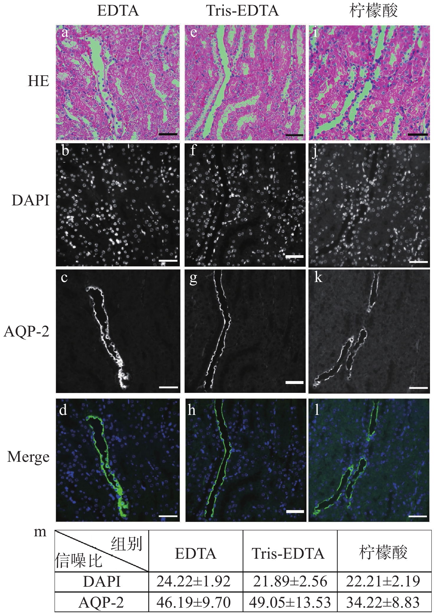
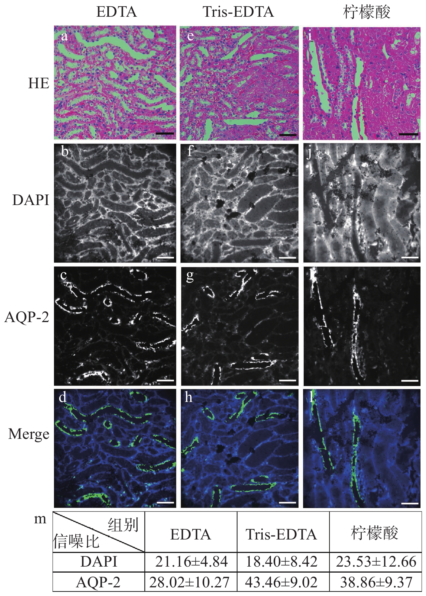
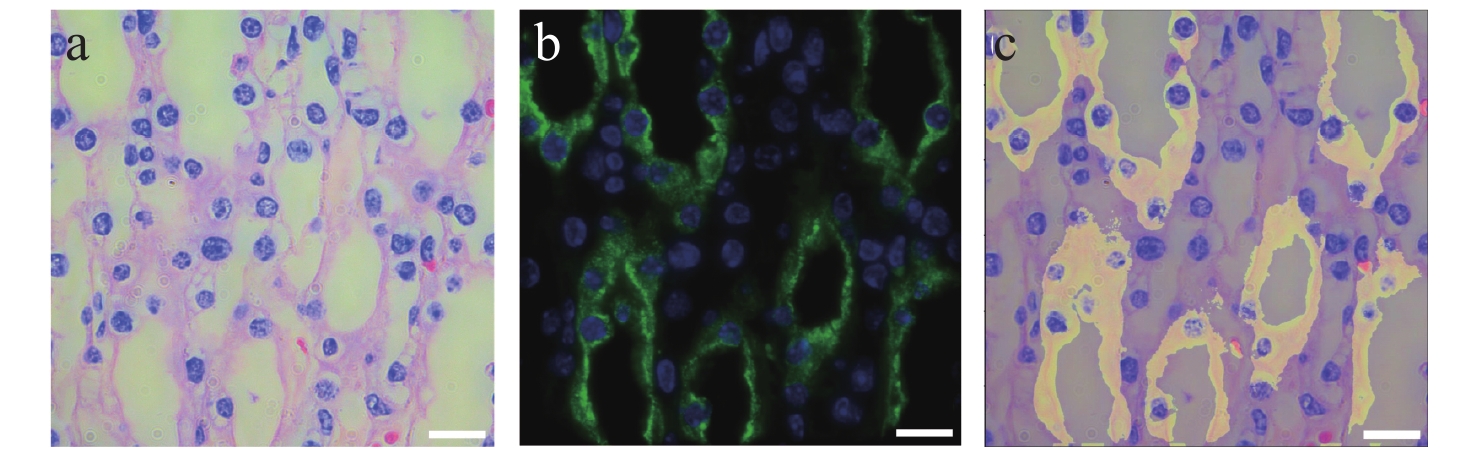
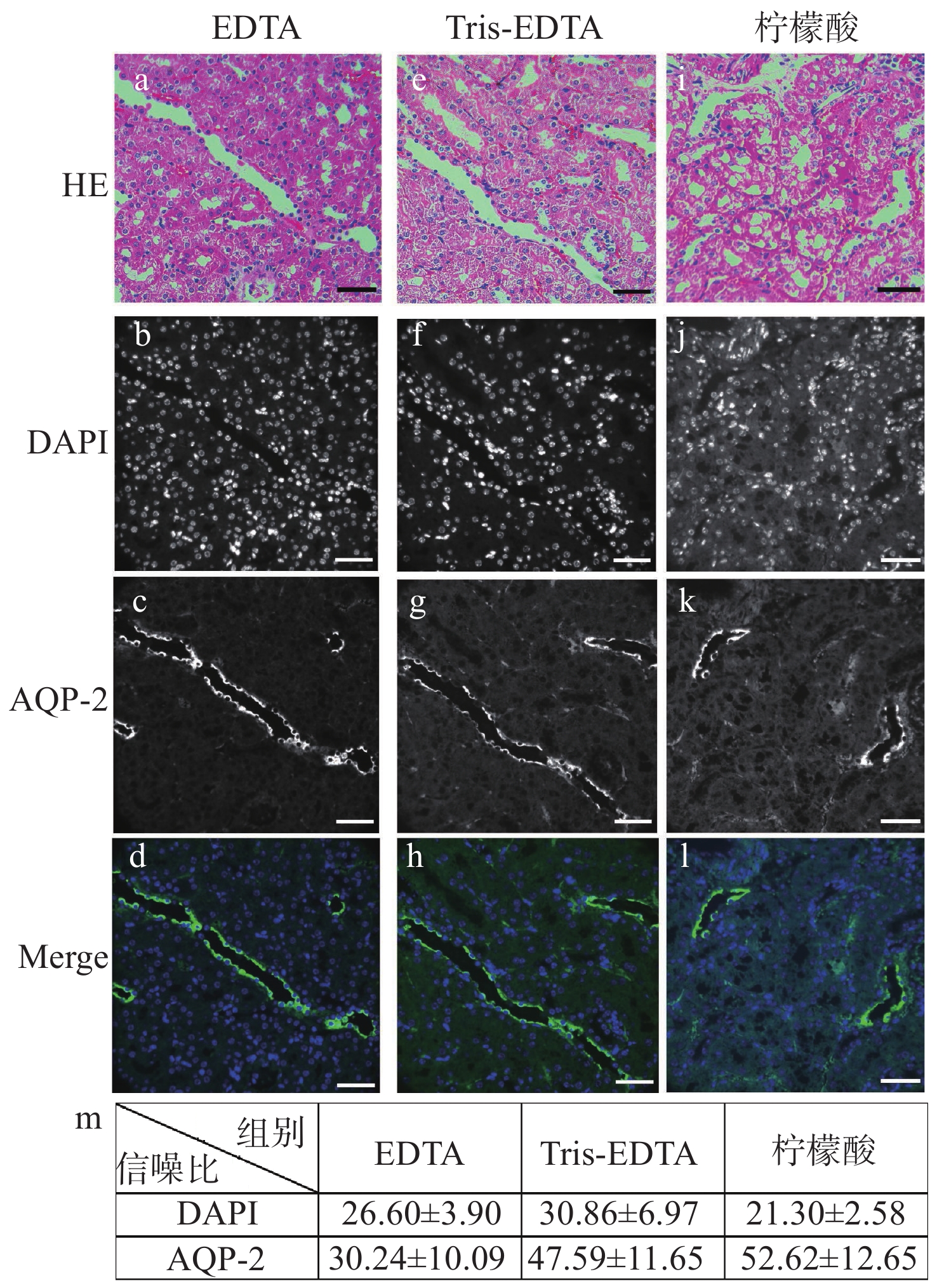
Bright-field imaging can provide cellular and histological morphological information, while fluorescence imaging can provide expression information of key proteins. Dual-modal correlation imaging based on both techniques is currently a common method for examining tissue samples in medical and scientific research. In clinical examination, however, correlation imaging between adjacent tissue slices is often used for observation. In such cases, both the tissue structure and the cellular level may be altered more or less, which is unfavorable when the sample volume is insufficient, the number of cells on the slices is limited, or precise point-to-point morphological information is required. Therefore, the development of single-slice dual-modal optical correlation imaging techniques which provides both tissue morphology and the distribution and expression of multiple target proteins on a single slice, can help to more accurately describe tumors and their microenvironment. This technique is particularly important in renal pathological testing where sample size is small. Renal pathology requires the use of bright-field imaging to obtain pathomorphological information of tissues and cells after hematoxylin-eosin staining, while the use of fluorescence imaging to obtain the distribution and expression of multiple target proteins is a mandatory molecular test for renal pathology screening. This paper focuses on the tissue sample processing methods that allow the coexistence of hematoxylin-eosin staining and immunofluorescence staining on the same renal slice. Improvements and comparative evaluations of the staining, de-colorizing and re-staining processes, as well as innovative fusion techniques for single-slice dual-modal imaging.

| [1] |
MASOOD S. The changing role of pathologists from morphologists to molecular pathologists in the era of precision medicine[J]. The Breast Journal, 2020, 26(1): 27-34. doi: 10.1111/tbj.13728
|
| [2] |
王义强, 林方睿, 胡睿, 等. 大视场光学显微成像技术[J]. 中国光学,2022,15(6):1194-1210. doi: 10.37188/CO.2022-0098
WANG Y Q, LIN F R, HU R, et al. Large field-of-view optical microscopic imaging technology[J]. Chinese Optics, 2022, 15(6): 1194-1210. (in Chinese) doi: 10.37188/CO.2022-0098
|
| [3] |
王鹏, 周瑶, 赵宇轩, 等. 用于多尺度高分辨率三维成像的双环光片荧光显微技术[J]. 中国光学,2022,15(6):1321-1331.
WANG P, ZHOU Y, ZHAO Y X, et al. Double-ring-modulated light sheet fluorescence microscopic technique for multi-scale high-resolution 3D imaging[J]. Chinese Optics, 2022, 15(6): 1321-1331. (in Chinese)
|
| [4] |
HARMS P W, FRANKEL T L, MOUTAFI M, et al. Multiplex immunohistochemistry and immunofluorescence: a practical update for pathologists[J]. Modern Pathology, 2023, 36(7): 100197. doi: 10.1016/j.modpat.2023.100197
|
| [5] |
LIM H G, LIU H C, YOON C W, et al. Investigation of cell mechanics using single-beam acoustic tweezers as a versatile tool for the diagnosis and treatment of highly invasive breast cancer cell lines: an in vitro study[J]. Microsystems & Nanoengineering, 2020, 6: 39.
|
| [6] |
MCNAMARA K K, KALMAR J R. Pearls and pitfalls in the diagnosis of small oral biopsies[J]. Seminars in Diagnostic Pathology, 2023, 40(5): 313-320. doi: 10.1053/j.semdp.2023.03.001
|
| [7] |
LÜTGERATH C, WEIß C, BÖER-AUER A. Clinicopathological features and histological tumor residues in re-excision specimens of incompletely resected basal cell carcinomas[J]. JDDG:Journal der Deutschen Dermatologischen Gesellschaft, 2022, 20(11): 1476-1483.
|
| [8] |
MORRISON L E, LEFEVER M R, LEWIS H N, et al. Conventional histological and cytological staining with simultaneous immunohistochemistry enabled by invisible chromogens[J]. Laboratory Investigation, 2022, 102(5): 545-553. doi: 10.1038/s41374-021-00714-2
|
| [9] |
FENG CH Y, LIU F. Artificial intelligence in renal pathology: current status and future[J]. Biomolecules and Biomedicine, 2023, 23(2): 225-234.
|
| [10] |
WALKER P D, CAVALLO T, BONSIB S M. Practice guidelines for the renal biopsy[J]. Modern Pathology, 2004, 17(12): 1555-1563. doi: 10.1038/modpathol.3800239
|
| [11] |
OZAWA A, SAKAUE M. New decolorization method produces more information from tissue sections stained with hematoxylin and eosin stain and masson-trichrome stain[J]. Annals of Anatomy-Anatomischer Anzeiger, 2020, 227: 151431. doi: 10.1016/j.aanat.2019.151431
|
| [12] |
FRANCIS R J, FERGUSON D, KEMPSTER S, et al. Blood identified and quantified in formalin fixed paraffin embedded lung sections using eosin fluorescence[J]. Histochemistry and Cell Biology, 2022, 158(4): 383-388. doi: 10.1007/s00418-022-02130-z
|
| [13] |
李丽, 杨桂芳. HE染色切片褪色后再进行免疫组化染色方法的比较[J]. 数理医药学杂志,2015,28(11):1618-1619. doi: 10.3969/j.issn.1004-4337.2015.11.015
LI L, YANG G F. Comparison of immunohistochemical staining methods for HE stained sections after de-colorizing[J]. Journal of Mathematical Medicine and Pharmacy, 2015, 28(11): 1618-1619. (in Chinese) (查阅网上资料, 未找到对应的英文翻译, 请确认) . doi: 10.3969/j.issn.1004-4337.2015.11.015
|
| [14] |
王兴波, 陈怀敏, 王秀珍, 等. HE染色褪色后免疫染色的病理观察[J]. 中国医药导报, 2007, 4(22): 124, 157.
WANG X B, CHEN H M, WANG X ZH, et al. Pathological observation of immunostaining after HE staining de-colorizing[J]. Chinese Medical Bulletin, 2007, 4(22): 124, 157. (in Chinese) (查阅网上资料, 未找到对应的英文翻译, 请确认) .
|
| [15] |
梁龄尹, 朱小兰, 骆新兰. 小标本切片HE染色褪色后再进行4种特殊染色方法的探讨[J]. 诊断病理学杂志,2020,27(6):443-444. doi: 10.3969/j.issn.1007-8096.2020.06.020
LIANG L Y, ZHU X L, LUO X L. Discussion on four special staining methods after HE staining de-colorizing of small specimen sections[J]. Chinese Journal of Diagnostic Pathology, 2020, 27(6): 443-444. (in Chinese) (查阅网上资料, 未找到对应的英文翻译, 请确认). doi: 10.3969/j.issn.1007-8096.2020.06.020
|
| [16] |
蒋绍仟. HE褪色后免疫组化染色防脱片方法的应用[J]. 四川肿瘤防治,2000(3):176.
JANG SH Q. Application of immunohistochemical staining anti-stripping method after HE de-colorizing[J]. Cancer Prevention and Treatment in Sichuan, 2000(3): 176. (in Chinese) (查阅网上资料, 未找到对应的英文翻译, 请确认) .
|
| [17] |
刘海芳. HE染色切片褪色后免疫组化染色方法研究[J]. 中国现代医生,2011,49(17):81-82. doi: 10.3969/j.issn.1673-9701.2011.17.039
LIU H F. Explore the method of immunohistochemiscal staining for HE slides after decoloration[J]. China Modern Doctor, 2011, 49(17): 81-82. (in Chinese) doi: 10.3969/j.issn.1673-9701.2011.17.039
|
| [18] |
李钦丽, 张继伟. HE切片经不同方法褪色后行EGFR基因突变检测的对比分析[J]. 临床与实验病理学杂志,2021,37(8):1004-1006.
LI Q L, ZHANG J W. Comparative analysis of EGFR gene mutation detection after different Methods of de-colorizing HE slices[J]. Chinese Journal of Clinical and Experimental Pathology, 2021, 37(8): 1004-1006. (in Chinese) (查阅网上资料, 未找到对应的英文翻译, 请确认) .
|
| [19] |
章克萍, 龙飞. 组织苏木精-伊红染色的石蜡切片褪色后还原对比染色[J]. 实用临床医学,2008,9(1):16. doi: 10.3969/j.issn.1009-8194.2008.01.005
ZHANG K P, LONG F. Reduction contrast staining after de-colorizing of paraffin slices stained with hematoxylin eosin in tissue[J]. Practical Clinical Medicine, 2008, 9(1): 16. (in Chinese) (查阅网上资料, 未找到对应的英文翻译, 请确认) . doi: 10.3969/j.issn.1009-8194.2008.01.005
|
| [20] |
高洪彬, 梁十, 郭扬清, 等. HE切片褪色后进行免疫荧光染色的方法探讨[J]. 临床与实验病理学杂志,2020,36(10):1241-1242. doi: 10.13315/j.cnki.cjcep.2020.10.027
GAO H B, LIANG SH, GUO Y Q, et al. Discussion on the method of immunofluorescence staining after HE section de-colorizing[J]. Chinese Journal of Clinical and Experimental Pathology, 2020, 36(10): 1241-1242. (in Chinese) (查阅网上资料, 未找到对应的英文翻译, 请确认) . doi: 10.13315/j.cnki.cjcep.2020.10.027
|
| [21] |
KOMURA D, ONOYAMA T, SHINBO K, et al. Restaining-based annotation for cancer histology segmentation to overcome annotation-related limitations among pathologists[J]. Patterns, 2023, 4(2): 100688. doi: 10.1016/j.patter.2023.100688
|
| [22] |
LI ZH M, MUENCH G, GOEBEL S, et al. Flow chamber staining modality for real-time inspection of dynamic phenotypes in multiple histological stains[J]. PLoS One, 2023, 18(5): e0284444. doi: 10.1371/journal.pone.0284444
|
| [23] |
JOHANN D J, SHIN I J, ROBERGE A, et al. Effect of antigen retrieval on genomic DNA from immunodissected samples[J]. Journal of Histochemistry & Cytochemistry, 2022, 70(9): 643-658.
|
| [24] |
GEORGE B, HAQUE A, SAHU V, et al. Enhancing antigen retrieval to unmask signaling phosphoproteins in formalin-fixed archival tissues[J]. Applied Immunohistochemistry & Molecular Morphology, 2022, 30(5): 333-339.
|
| [25] |
DUNKENBERGER L, DEL VALLE L. Antigen retrieval and signal amplification[M]//DEL VALLE L. Immunohistochemistry and Immunocytochemistry: Methods and Protocols. New York: Humana, 2022: 65-74.
|
Figures(4)

 DownLoad: