| Citation: | ZHANG Zhi-miao, WANG Cheng-miao, XIE Mian, LIN Yu, HAN Ye-ming, DENG Yong-bo, GUO Chang-liang, FU Qiang. Design of miniature head-mounted fluorescence microscope based on metalens[J]. Chinese Optics. doi: 10.37188/CO.2023-0237

|
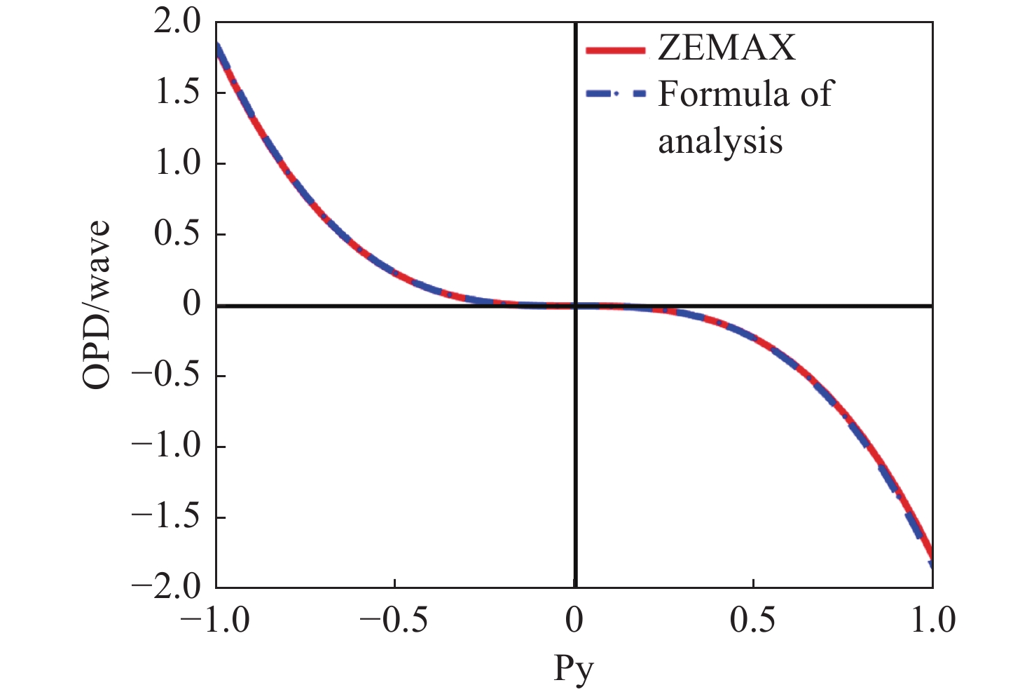
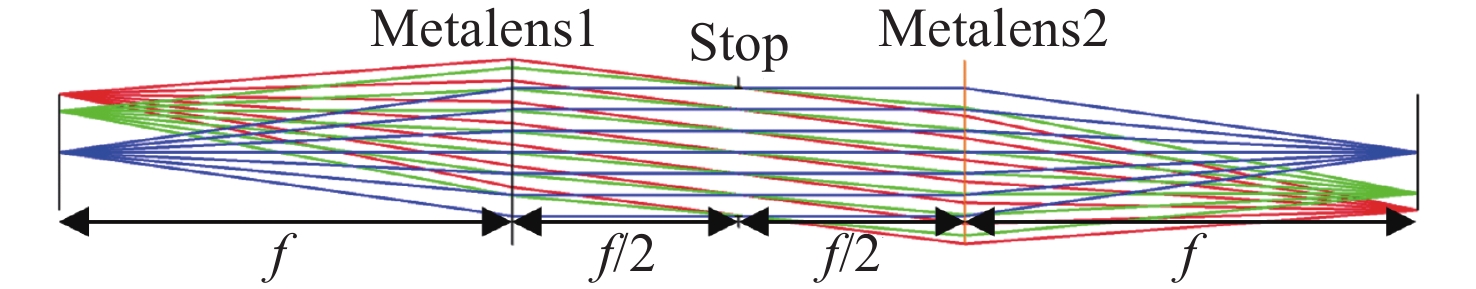
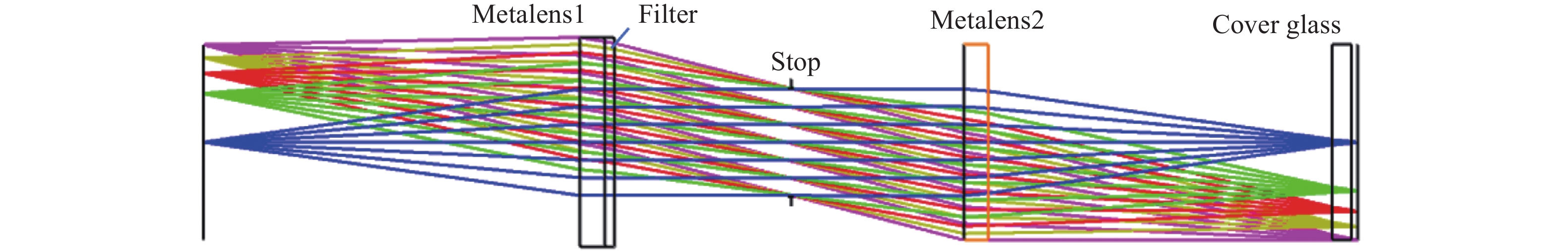
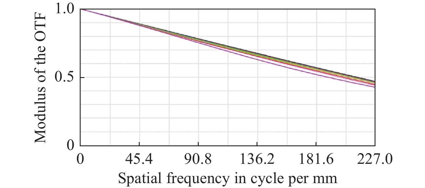
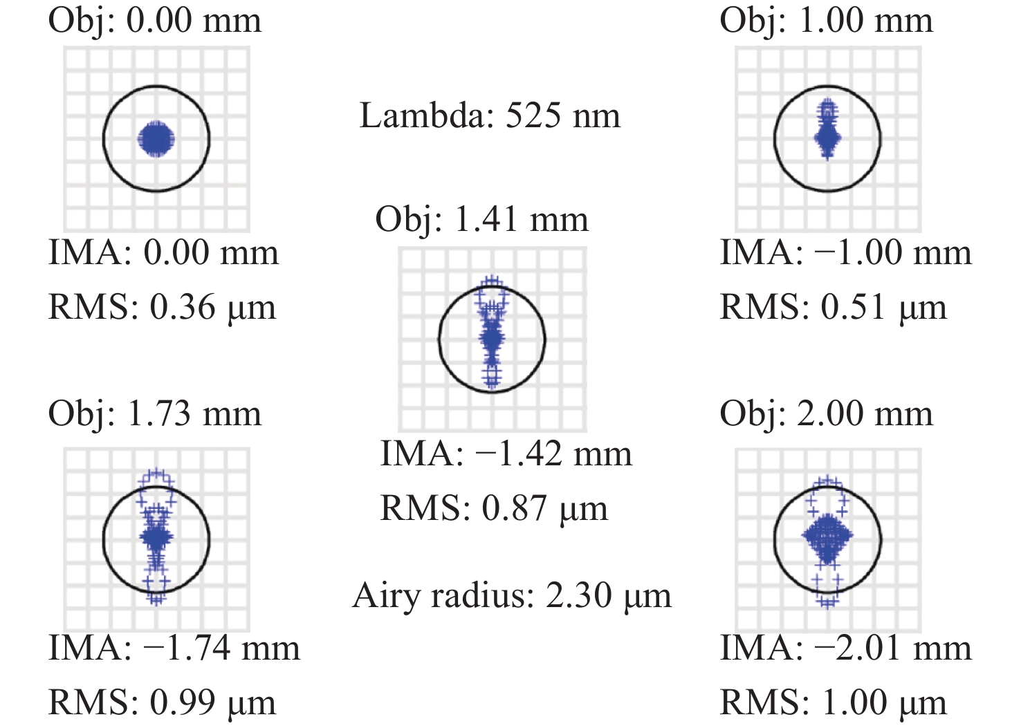
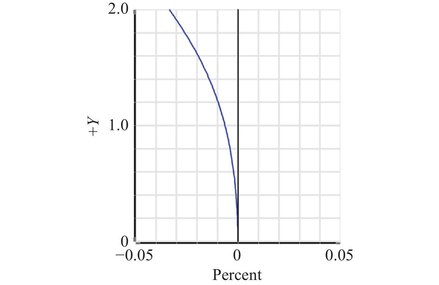
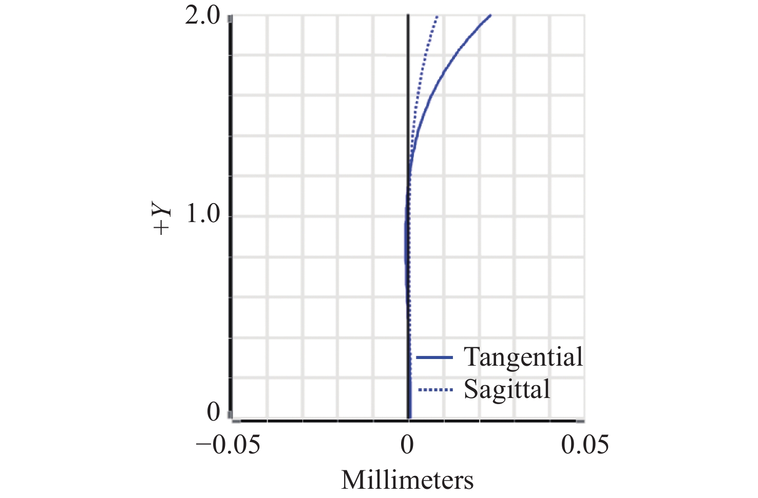
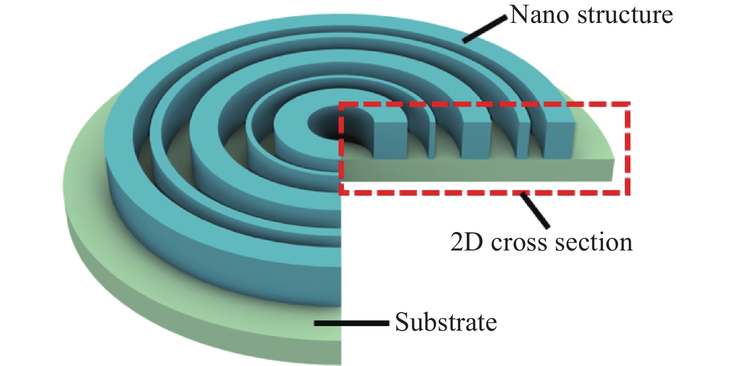
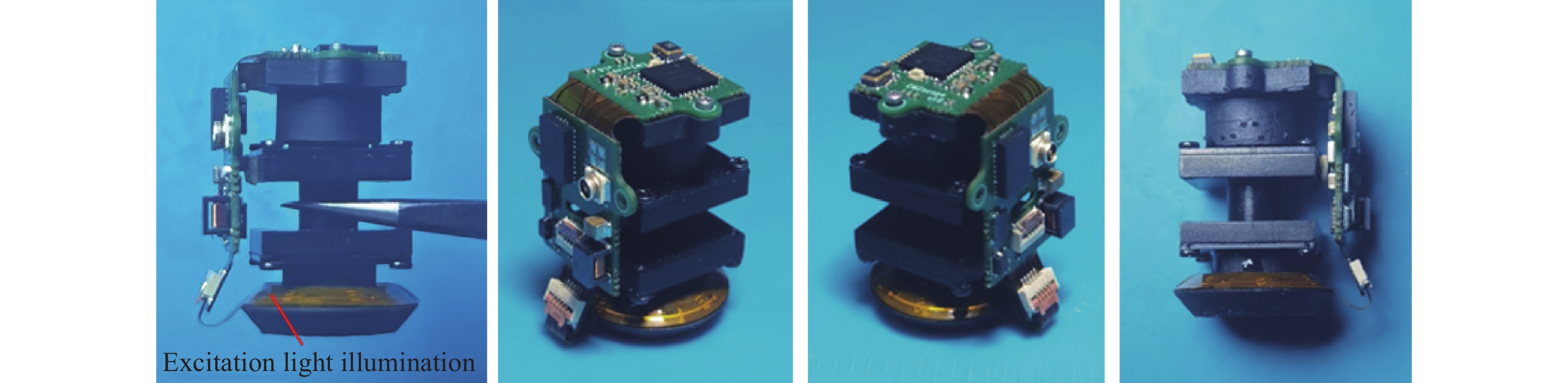
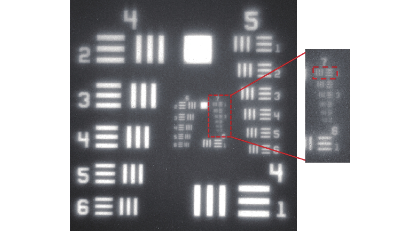
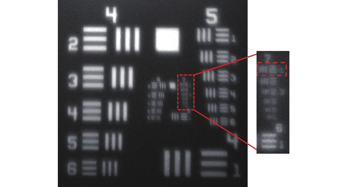
The recent advent of miniature head-mounted fluorescence microscopes has revolutionized brain science research, enabling real-time imaging of neural activity in the brains of free-moving animals. However, the pursuit of miniaturization and reduced weight often results in a limited field of view, constraining the number of neurons observable. While larger field-of-view systems exist, their increased weight can impede the natural behaviors of the subjects. Addressing these limitations, a novel design utilizing a metalens schematic is proposed. This approach offers the benefits of being ultra-light, ultra-thin, and capable of high-quality imaging. By deriving the aberration formula specific to hyperbolic phase metalens and using it as a foundation, a design for a miniature fluorescence microscope was developed. This microscope boasts a 4 mm×4 mm field of view and a numerical aperture (NA) of 0.14, effectively correcting seven primary aberrations. The resulting prototype, weighing a mere 4.11 g, achieves a resolution of 7.8 μm across the entire field of view. This performance is sufficient to image neural activity in the brains of freely moving mice with single-cell resolution.

| [1] |
GRIENBERGER C, KONNERTH A. Imaging calcium in neurons[J]. Neuron, 2012, 73(5): 862-885. doi: 10.1016/j.neuron.2012.02.011
|
| [2] |
YU H, SENARATHNA J, TYLER B M, et al. Miniaturized optical neuroimaging in unrestrained animals[J]. NeuroImage, 2015, 113: 397-406. doi: 10.1016/j.neuroimage.2015.02.070
|
| [3] |
CHEN SH Y, WANG Z CH, ZHANG D, et al. Miniature fluorescence microscopy for imaging brain activity in freely-behaving animals[J]. Neuroscience Bulletin, 2020, 36(10): 1182-1190. doi: 10.1007/s12264-020-00561-z
|
| [4] |
付强, 张智淼, 赵尚男, 等. 微型头戴式单光子荧光显微成像技术研究进展[J]. 中国光学,2023,16(5):1010-1021. doi: 10.37188/CO.2023-0007
FU Q, ZHANG ZH M, ZHAO SH N, et al. Research progress of miniature head-mounted single photon fluorescence microscopic imaging technique[J]. Chinese Optics, 2023, 16(5): 1010-1021. (in Chinese). doi: 10.37188/CO.2023-0007
|
| [5] |
RYNES M L, SURINACH D A, LINN S, et al. Miniaturized head-mounted microscope for whole-cortex mesoscale imaging in freely behaving mice[J]. Nature Methods, 2021, 18(4): 417-425. doi: 10.1038/s41592-021-01104-8
|
| [6] |
GHOSH K K, BURNS L D, COCKER E D, et al. Miniaturized integration of a fluorescence microscope[J]. Nature Methods, 2011, 8(10): 871-878. doi: 10.1038/nmeth.1694
|
| [7] |
CAI D J, AHARONI D, SHUMAN T, et al. A shared neural ensemble links distinct contextual memories encoded close in time[J]. Nature, 2016, 534(7605): 115-118. doi: 10.1038/nature17955
|
| [8] |
LIBERTI W A, PERKINS L N, LEMAN D P, et al. An open source, wireless capable miniature microscope system[J]. Journal of Neural Engineering, 2017, 14(4): 045001. doi: 10.1088/1741-2552/aa6806
|
| [9] |
SKOCEK O, NÖBAUER T, WEILGUNY L, et al. High-speed volumetric imaging of neuronal activity in freely moving rodents[J]. Nature Methods, 2018, 15(6): 429-432. doi: 10.1038/s41592-018-0008-0
|
| [10] |
JACOB A D, RAMSARAN A I, MOCLE A J, et al. A compact head-mounted endoscope for in vivo calcium imaging in freely behaving mice[J]. Current Protocols in Neuroscience, 2018, 84(1): e51. doi: 10.1002/cpns.51
|
| [11] |
AHARONI D, KHAKH B S, SILVA A J, et al. All the light that we can see: a new era in miniaturized microscopy[J]. Nature Methods, 2019, 16(1): 11-13. doi: 10.1038/s41592-018-0266-x
|
| [12] |
BAGRAMYAN A. Lightweight 1-photon miniscope for imaging in freely behaving animals at subcellular resolution[J]. IEEE Photonics Technology Letters, 2020, 32(15): 909-912. doi: 10.1109/LPT.2020.3004283
|
| [13] |
DE GROOT A, VAN DEN BOOM B J G, VAN GENDEREN R M, et al. NINscope, a versatile miniscope for multi-region circuit investigations[J]. eLife, 2020, 9: e49987. doi: 10.7554/eLife.49987
|
| [14] |
YANNY K, ANTIPA N, LIBERTI W, et al. Miniscope3D: optimized single-shot miniature 3D fluorescence microscopy[J]. Light:Science & Applications, 2020, 9: 171.
|
| [15] |
SHUMAN T, AHARONI D, CAI D J, et al. Breakdown of spatial coding and interneuron synchronization in epileptic mice[J]. Nature Neuroscience, 2020, 23(2): 229-238. doi: 10.1038/s41593-019-0559-0
|
| [16] |
BAGRAMYAN A, TABOURIN L, RASTQAR A, et al. Focus-tunable microscope for imaging small neuronal processes in freely moving animals[J]. Photonics Research, 2021, 9(7): 1300. doi: 10.1364/PRJ.418154
|
| [17] |
WANG Y ZH, MA ZH T, LI W ZH, et al. Cable-free brain imaging for multiple free-moving animals with miniature wireless microscopes[J]. Journal of Biomedical Optics, 2023, 28(2): 026503.
|
| [18] |
SCOTT B B, THIBERGE S Y, GUO C Y, et al. Imaging cortical dynamics in GCaMP transgenic rats with a head-mounted widefield macroscope[J]. Neuron, 2018, 100(5): 1045-1058. e5.
|
| [19] |
GUO CH L, BLAIR G J, SEHGAL M, et al. Miniscope-LFOV: a large-field-of-view, single-cell-resolution, miniature microscope for wired and wire-free imaging of neural dynamics in freely behaving animals[J]. Science Advances, 2023, 9(16): 3918-3918. doi: 10.1126/sciadv.adg3918
|
| [20] |
XU B B, LI H M, GAO SH L, et al. Metalens-integrated compact imaging devices for wide-field microscopy[J]. Advanced Photonics, 2020, 2(6): 066004.
|
| [21] |
TSENG E, COLBURN S, WHITEHEAD J, et al. Neural Nano-optics for high-quality thin lens imaging[J]. Nature Communications, 2021, 12(1): 6493. doi: 10.1038/s41467-021-26443-0
|
| [22] |
LIU Y, YU Q Y, CHEN Z M, et al. Meta-objective with sub-micrometer resolution for microendoscopes[J]. Photonics Research, 2021, 9(2): 106-115. doi: 10.1364/PRJ.406197
|
| [23] |
AIETA F, GENEVET P, KATS M, et al. Aberrations of flat lenses and aplanatic metasurfaces[J]. Optics Express, 2013, 21(25): 31530-31539. doi: 10.1364/OE.21.031530
|
| [24] |
YOUNG M. Zone plates and their aberrations[J]. Journal of the Optical Society of America, 1972, 62(8): 972-976. doi: 10.1364/JOSA.62.000972
|
| [25] |
GROSS H. Handbook of Optical Systems (Volume 3: Aberration Theory and Correction of Optical Systems)[M]. Weinheim: John Wiley & Sons Inc, 2007.
|
| [26] |
WANG CH M, LIN Y, HAN Y M, et al. Fabricable concentric-ring metalens with high focusing efficiency based on two-dimensional subwavelength unit splicing[J]. Optics Express, 2023, 31(20): 33596-33607. doi: 10.1364/OE.500688
|
| [27] |
JIN ZH, LIN Y, WANG CH M, et al. Topologically optimized concentric-nanoring metalens with 1 mm diameter, 0.8 NA and 600 nm imaging resolution in the visible[J]. Optics Express, 2023, 31(6): 10489-10499. doi: 10.1364/OE.478680
|
Figures(18) / Tables(2)

 DownLoad: