| Citation: | ZHOU Wen-chao, LI Zheng-hao, WU Jie. Research progress of single molecule biological detection methods and applications[J]. Chinese Optics, 2022, 15(5): 878-894. doi: 10.37188/CO.2022-0129

|
Single molecule biological detection technology is an efficient technology to understand the dynamic characteristics of various biomolecules at the single molecule level and explore their structure and function. The advantage of this technology is that it can detect the heterogeneity of free energy on a single molecule, which is beyond the traditional methods. Therefore, researchers use it to solve long-standing problems in complex biological systems, heterogeneous catalysis, biomolecular interactions, enzyme systems and conformational changes. In terms of medical detection, detecting specific information about single molecules or their interactions with biological factors is not only crucial for the early diagnosis and treatment of various diseases such as cancer, but also has great potential for real-time detection and precision medicine. The advantages of high specificity and high precision of single-molecule bioassays are used to real-time detection of single biomolecules in molecular populations, and can be combined with multiple high-throughput analysis for the precise diagnosis of clinical samples. In this paper, the principle of single molecule detection and the application of biosensing are introduced, and the detection methods and related applications are summarized. Finally, the prospect and development direction of this research direction are discussed.

| [1] |
HALL C E. Method for the observation of macromolecules with the electron microscope illustrated with micrographs of DNA[J]. The Journal of Biophysical and Biochemical Cytology, 1956, 2(5): 625-628. doi: 10.1083/jcb.2.5.625
|
| [2] |
ROTMAN B. Measurement of activity of single molecules of β -D-galactosidase[J]. Proceedings of the National Academy of Sciences of the United States of America, 1961, 47(12): 1981-1991. doi: 10.1073/pnas.47.12.1981
|
| [3] |
MILLER H, ZHOU ZH K, SHEPHERD J, et al. Single-molecule techniques in biophysics: a review of the progress in methods and applications[J]. Reports on Progress in Physics, 2018, 81(2): 024601. doi: 10.1088/1361-6633/aa8a02
|
| [4] |
HIRSCHFELD T. Optical microscopic observation of single small molecules[J]. Applied Optics, 1976, 15(12): 2965-2966. doi: 10.1364/AO.15.002965
|
| [5] |
GELLES J, SCHNAPP B J, SHEETZ M P. Tracking kinesin-driven movements with nanometre-scale precision[J]. Nature, 1988, 331(6155): 450-453. doi: 10.1038/331450a0
|
| [6] |
ORRIT M, BERNARD J. Single pentacene molecules detected by fluorescence excitation in a p-terphenyl crystal[J]. Physical Review Letters, 1990, 65(21): 2716-2719. doi: 10.1103/PhysRevLett.65.2716
|
| [7] |
KNEIPP K, WANG Y, KNEIPP H, et al. Single molecule detection using surface-enhanced Raman scattering (SERS)[J]. Physical Review Letters, 1997, 78(9): 1667-1670. doi: 10.1103/PhysRevLett.78.1667
|
| [8] |
LU H P, XUN L, XIE X S. Single molecule enzymatic dynamics[J]. Science, 1998, 282(5395): 1877-1882.
|
| [9] |
VOGELSTEIN B, KINZLER K W. Digital PCR[J]. Proceedings of the National Academy of Sciences of the United States of America, 1999, 96(16): 9236-9241. doi: 10.1073/pnas.96.16.9236
|
| [10] |
KOREN S, SCHATZ M C, WALENZ B P, et al. Hybrid error correction and de novo assembly of single-molecule sequencing reads[J]. Nature Biotechnology, 2012, 30(7): 693-700. doi: 10.1038/nbt.2280
|
| [11] |
GOOTENBERG J S, ABUDAYYEH O O, LEE J W, et al. Nucleic acid detection with CRISPR-Cas13a/C2c2[J]. Science, 2017, 356(6336): 438-442. doi: 10.1126/science.aam9321
|
| [12] |
FUXREITER M, VENDRUSCOLO M. Generic nature of the condensed states of proteins[J]. Nature Cell Biology, 2021, 23(6): 587-594. doi: 10.1038/s41556-021-00697-8
|
| [13] |
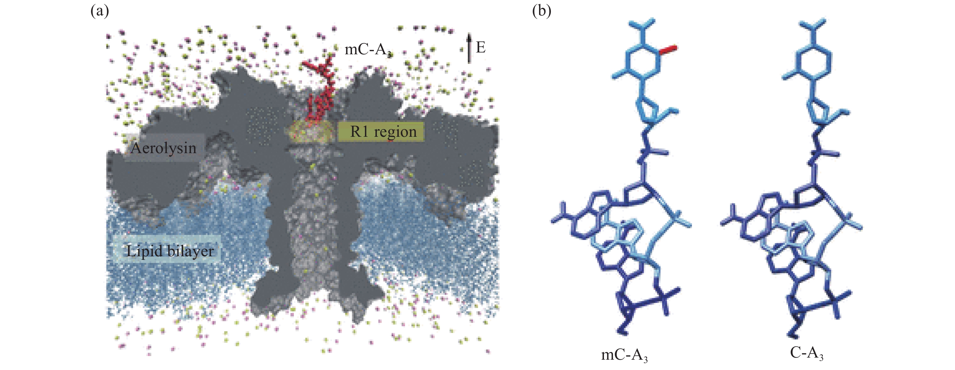
WANG Y Q, GUAN X Y, ZHANG S Y, et al. Structural-profiling of low molecular weight RNAs by nanopore trapping/translocation using Mycobacterium smegmatis porin A[J]. Nature Communications, 2021, 12(1): 3368. doi: 10.1038/s41467-021-23764-y
|
| [14] |
RUAN J B, XIA SH Y, LIU X, et al. Cryo-EM structure of the gasdermin A3 membrane pore[J]. Nature, 2018, 557(7703): 62-67. doi: 10.1038/s41586-018-0058-6
|
| [15] |
WANG J Z, HU M J, WANG J, et al. Reconstitution and structure of a plant NLR resistosome conferring immunity[J]. Science, 2019, 364(6435): eaav5870. doi: 10.1126/science.aav5870
|
| [16] |
LANGECKER M, ARNAUT V, MARTIN T G, et al. Synthetic lipid membrane channels formed by designed DNA nanostructures[J]. Science, 2012, 338(6109): 932-936. doi: 10.1126/science.1225624
|
| [17] |
KASIANOWICZ J J, BRANDIN E, BRANTON D. Characterization of individual polynucleotide molecules using a membrane channel[J]. Proceedings of the National Academy of Sciences of the United States of America, 1996, 93(24): 13770-13773. doi: 10.1073/pnas.93.24.13770
|
| [18] |
CHAVIS A E, BRADY K T, HATMAKER G A, et al. Single molecule nanopore spectrometry for peptide detection[J]. ACS Sensors, 2017, 2(9): 1319-1328. doi: 10.1021/acssensors.7b00362
|
| [19] |
RAHMAN M M, SAMPAD M J N, HAWKINS A, et al. Recent advances in integrated solid-state nanopore sensors[J]. Lab on a Chip, 2021, 21(16): 3030-3052. doi: 10.1039/D1LC00294E
|
| [20] |
MACCAFERRIN, BARBILLON G, KOYAAN, et al. Recent advances in plasmonic nanocavities for single-molecule spectroscopy[J]. Nanoscale Advance, 2021, 3(3): 633-642.
|
| [21] |
ASSAD O N, GILBOA T, SPITZBERG J, et al. Light-enhancing plasmonic-nanopore biosensor for superior single-molecule detection[J]. Advanced Materials, 2017, 29(9): 1605442. doi: 10.1002/adma.201605442
|
| [22] |
BUCHFINK B, REUTER K, DROST H G. Sensitive protein alignments at tree-of-life scale using DIAMOND[J]. Nature Methods, 2021, 18(4): 366-368. doi: 10.1038/s41592-021-01101-x
|
| [23] |
BELLENGUEZ C, KÜÇÜKALI F, JANSEN I E, et al. New insights into the genetic etiology of Alzheimer's disease and related dementias[J]. Nature Genetics, 2022, 54(4): 412-436. doi: 10.1038/s41588-022-01024-z
|
| [24] |
DEVESON I W, GONG B SH, LAI K, et al. Evaluating the analytical validity of circulating tumor DNA sequencing assays for precision oncology[J]. Nature Biotechnology, 2021, 39(9): 1115-1128. doi: 10.1038/s41587-021-00857-z
|
| [25] |
BRINKERHOFF H, KANG A S W, LIU J Q, et al.. Infinite re-reading of single proteins at single-amino-acid resolution using nanopore sequencing[J]. BioRxiv, 2021, doi: 10.1126/science.abl4381.
|
| [26] |
SEN P, GUPTA M. Single nucleotide detection using bilayer MoS2 nanopores with high efficiency[J]. RSC Advances, 2021, 11(11): 6114-6123. doi: 10.1039/D0RA10222A
|
| [27] |
YAMAZAKI H, HU R, ZHAO Q, et al. Photothermally assisted thinning of silicon nitride membranes for ultrathin asymmetric nanopores[J]. ACS Nano, 2018, 12(12): 12472-12481. doi: 10.1021/acsnano.8b06805
|
| [28] |
BURCK N, GILBOA T, GADI A, et al. Nanopore identification of single nucleotide mutations in circulating tumor DNA by multiplexed ligation[J]. Clinical Chemistry, 2021, 67(5): 753-762. doi: 10.1093/clinchem/hvaa328
|
| [29] |
YUAN B, LI SH, YING Y L, et al. The analysis of single cysteine molecules with an aerolysin nanopore[J]. Analyst, 2020, 145(4): 1179-1183. doi: 10.1039/C9AN01965K
|
| [30] |
PIGUET F, OULDALI H, PASTORIZA-GALLEGO M, et al. Identification of single amino acid differences in uniformly charged homopolymeric peptides with aerolysin nanopore[J]. Nature Communications, 2018, 9(1): 966. doi: 10.1038/s41467-018-03418-2
|
| [31] |
CAO C, CIRAUQUI N, MARCAIDA M J, et al. Single-molecule sensing of peptides and nucleic acids by engineered aerolysin nanopores[J]. Nature communications, 2019, 10(1): 1-11.
|
| [32] |
ZHANG SH L, HUANG G, VERSLOOT R C A, et al. Bottom-up fabrication of a proteasome–nanopore that unravels and processes single proteins[J]. Nature Chemistry, 2021, 13(12): 1192-1199. doi: 10.1038/s41557-021-00824-w
|
| [33] |
LI M Y, YING Y L, YU J, et al. Revisiting the origin of nanopore current blockage for volume difference sensing at the atomic level[J]. JACS Au, 2021, 1(7): 967-976. doi: 10.1021/jacsau.1c00109
|
| [34] |
LEIRS K, KUMAR P T, DECROP D, et al. Bioassay development for ultrasensitive detection of influenza a nucleoprotein using digital ELISA[J]. Analytical Chemistry, 2016, 88(17): 8450-8458. doi: 10.1021/acs.analchem.6b00502
|
| [35] |
SHAFAGH R Z, DECROP D, VEN K, et al. Reaction injection molding of hydrophilic-in-hydrophobic femtolitre-well arrays[J]. Microsystems &Nanoengineering, 2019, 5: 25.
|
| [36] |
RISSIN D M, WALT D R. Digital readout of target binding with attomole detection limits via enzyme amplification in femtoliter arrays[J]. Journal of the American Chemical Society, 2006, 128(19): 6286-6287. doi: 10.1021/ja058425e
|
| [37] |
RISSIN D M, KAN C W, CAMPBELL T G, et al. Single-molecule enzyme-linked immunosorbent assay detects serum proteins at subfemtomolar concentrations[J]. Nature Biotechnology, 2010, 28(6): 595-599. doi: 10.1038/nbt.1641
|
| [38] |
LEE J, CRAMPTON K T, TALLARIDA N, et al. Visualizing vibrational normal modes of a single molecule with atomically confined light[J]. Nature, 2019, 568(7750): 78-82. doi: 10.1038/s41586-019-1059-9
|
| [39] |
YERA H, OK V, KUET F L K, et al. PCR and culture for diagnosis of Acanthamoeba keratitis[J]. British Journal of Ophthalmology, 2021, 105(9): 1302-1306. doi: 10.1136/bjophthalmol-2020-316730
|
| [40] |
HINDSON B J, NESS K D, MASQUELIER D A, et al. High-throughput droplet digital PCR system for absolute quantitation of DNA copy number[J]. Analytical Chemistry, 2011, 83(22): 8604-8610. doi: 10.1021/ac202028g
|
| [41] |
CHEN Y J, QIAN CH, LIU CH ZH, et al. Nucleic acid amplification free biosensors for pathogen detection[J]. Biosensors and Bioelectronics, 2020, 153: 112049. doi: 10.1016/j.bios.2020.112049
|
| [42] |
GINES G, MENEZES R, NARA K, et al. Isothermal digital detection of microRNAs using background-free molecular circuit[J]. Science Advances, 2020, 6(4): eaay5952. doi: 10.1126/sciadv.aay5952
|
| [43] |
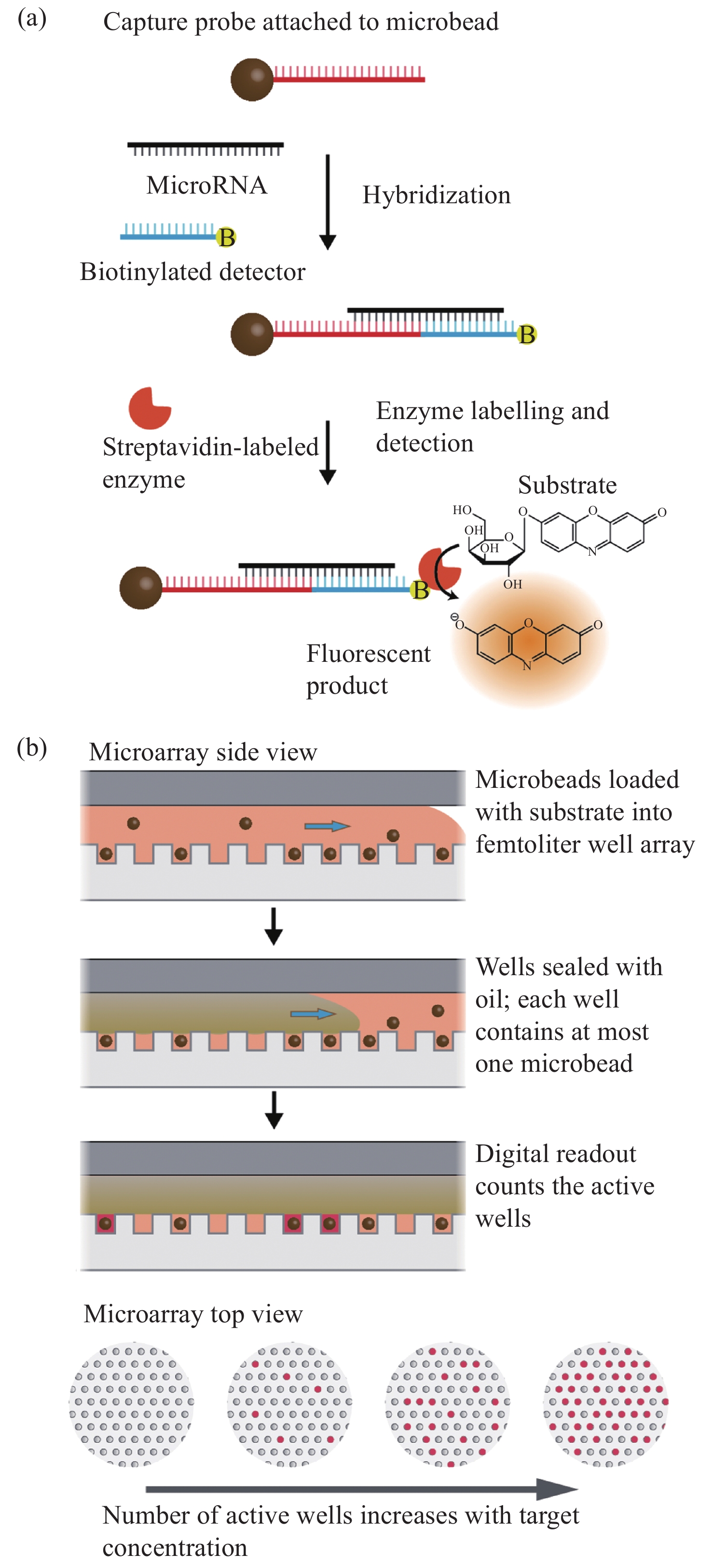
COHEN L, HARTMAN M R, AMARDEY-WELLINGTON A, et al. Digital direct detection of microRNAs using single molecule arrays[J]. Nucleic Acids Research, 2017, 45(14): e137. doi: 10.1093/nar/gkx542
|
| [44] |
ASHTON N J, LEUZY A, KARIKARI T K, et al. The validation status of blood biomarkers of amyloid and phospho-tau assessed with the 5-phase development framework for AD biomarkers[J]. European Journal of Nuclear Medicine and Molecular Imaging, 2021, 48(7): 2140-2156. doi: 10.1007/s00259-021-05253-y
|
| [45] |
GILL J, LATOUR L, DIAZ-ARRASTIA R, et al. Glial fibrillary acidic protein elevations relate to neuroimaging abnormalities after mild TBI[J]. Neurology, 2018, 91(15): e1385-e1389. doi: 10.1212/WNL.0000000000006321
|
| [46] |
THELIN E, AL NIMER F, FROSTELL A, et al. A serum protein biomarker panel improves outcome prediction in human traumatic brain injury[J]. Journal of Neurotrauma, 2019, 36(20): 2850-2862. doi: 10.1089/neu.2019.6375
|
| [47] |
PÉREZ-RUIZ E, DECROP D, VEN K, et al. Digital ELISA for the quantification of attomolar concentrations of Alzheimer's disease biomarker protein Tau in biological samples[J]. Analytica Chimica Acta, 2018, 1015: 74-81. doi: 10.1016/j.aca.2018.02.011
|
| [48] |
DINH T L, NGAN K C, SHOEMAKER C B, et al. Rapid and ultrasensitive detection of botulinum neurotoxin serotype A1 in human serum and urine using single-molecule array method[J]. Forensic Toxicology, 2017, 35(1): 179-184. doi: 10.1007/s11419-016-0336-7
|
| [49] |
WANG X, COHEN L, WANG J, et al. Competitive immunoassays for the detection of small molecules using single molecule arrays[J]. Journal of the American Chemical Society, 2018, 140(51): 18132-18139. doi: 10.1021/jacs.8b11185
|
| [50] |
KIM E, BAASKE M D, VOLLMER F. Towards next-generation label-free biosensors: recent advances in whispering gallery mode sensors[J]. Lab on a Chip, 2017, 17(7): 1190-1205. doi: 10.1039/C6LC01595F
|
| [51] |
SUBRAMANIAN S, VINCENT S, VOLLMER F. Effective linewidth shifts in single-molecule detection using optical whispering gallery modes[J]. Applied Physics Letters, 2020, 117(15): 151106. doi: 10.1063/5.0028113
|
| [52] |
SANTIAGO-CORDOBA M A, CETINKAYA M, BORISKINA S V, et al. Ultrasensitive detection of a protein by optical trapping in a photonic-plasmonic microcavity[J]. Journal of Biophotonics, 2012, 5(8-9): 629-638. doi: 10.1002/jbio.201200040
|
| [53] |
YU W Y, JIANG W C, LIN Q, et al. Cavity optomechanical spring sensing of single molecules[J]. Nature Communications, 2016, 7: 12311. doi: 10.1038/ncomms12311
|
| [54] |
BAILEY R C, WASHBURN A L, QAVI A J, et al. A robust silicon photonic platform for multiparameter biological analysis[J]. Proceedings of SPIE, 2009, 7220: 72200N. doi: 10.1117/12.809819
|
| [55] |
SHI H X, CUI J J, SULEMANA H, et al. Protein detection based on rolling circle amplification sensors[J]. Luminescence, 2021, 36(4): 842-848. doi: 10.1002/bio.4017
|
| [56] |
NITU F R, SAVU L, MURARU S, et al. Label-free homogeneous microRNA detection in cell culture medium based on graphene oxide and specific fluorescence quenching[J]. Nanomaterials, 2021, 11(2): 368. doi: 10.3390/nano11020368
|
| [57] |
VOLLMER F, ARNOLD S, KENG D. Single virus detection from the reactive shift of a whispering-gallery mode[J]. Proceedings of the National Academy of Sciences of the United States of America, 2008, 105(52): 20701-20704. doi: 10.1073/pnas.0808988106
|
| [58] |
CARDENOSA-RUBIO M C, ROBISON H M, BAILEY R C. Recent advances in environmental and clinical analysis using microring resonator–based sensors[J]. Current Opinion in Environmental Science &Health, 2019, 10: 38-46.
|
| [59] |
SHAO L B, JIANG X F, YU X CH, et al. Detection of single nanoparticles and lentiviruses using microcavity resonance broadening[J]. Advanced Materials, 2013, 25(39): 5616-5620. doi: 10.1002/adma201302572
|
| [60] |
DOMINGUEZ I, DEL VILLAR I, FUENTES O, et al. Dually nanocoated planar waveguides towards multi-parameter sensing[J]. Scientific Reports, 2021, 11(1): 3669. doi: 10.1038/s41598-021-83324-8
|
| [61] |
COGNETTI J S, STEINER D J, ABEDIN M, et al. Disposable photonics for cost-effective clinical bioassays: application to COVID-19 antibody testing[J]. Lab on a Chip, 2021, 21(15): 2913-2921. doi: 10.1039/D1LC00369K
|
| [62] |
ROBISON H M, ESCALANTE P, VALERA E, et al. Precision immunoprofiling to reveal diagnostic signatures for latent tuberculosis infection and reactivation risk stratification[J]. Integrative Biology, 2019, 11(1): 16-25. doi: 10.1093/intbio/zyz001
|
| [63] |
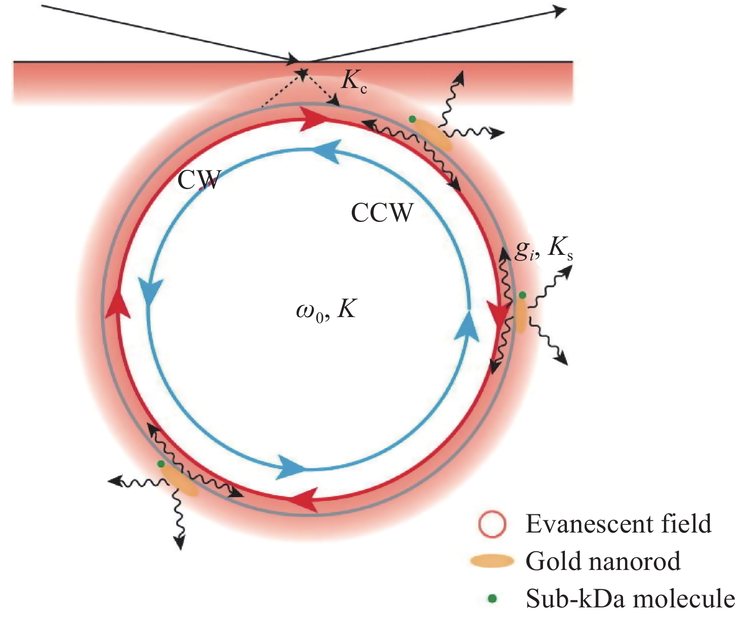
YU X CH, TANG SH J, LIU W J, et al. Single-molecule optofluidic microsensor with interface whispering gallery modes[J]. Proceedings of the National Academy of Sciences of the United States of America, 2022, 119(6): e2108678119. doi: 10.1073/pnas.2108678119
|
| [64] |
KIM E, BAASKE M D, VOLLMER F. In situ observation of single-molecule surface reactions from low to high affinities[J]. Advanced Materials, 2016, 28(45): 9941-9948. doi: 10.1002/adma.201603153
|
| [65] |
KIM E, BAASKE M D, SCHULDES I, et al. Label-free optical detection of single enzyme-reactant reactions and associated conformational changes[J]. Science Advances, 2017, 3(3): e1603044. doi: 10.1126/sciadv.1603044
|
| [66] |
SUBRAMANIAN S, JONES H B L, FRUSTACI S, et al. Sensing enzyme activation heat capacity at the single-molecule level using gold-nanorod-based optical whispering gallery modes[J]. ACS Applied Nano Materials, 2021, 4(5): 4576-4583. doi: 10.1021/acsanm.1c00176
|
| [67] |
AMBARTSUMYAN O, GRIBANYOV D, KUKUSHKIN V, et al. SERS-based biosensors for virus determination with oligonucleotides as recognition elements[J]. International Journal of Molecular Sciences, 2020, 21(9): 3373. doi: 10.3390/ijms21093373
|
| [68] |
FLEISCHMANN M, HENDRA P J, MCQUILLAN A J. Raman spectra of pyridine adsorbed at a silver electrode[J]. Chemical Physics Letters, 1974, 26(2): 163-166. doi: 10.1016/0009-2614(74)85388-1
|
| [69] |
LI W Y, CAMARGO P H C, LU X M, et al. Dimers of silver nanospheres: facile synthesis and their use as hot spots for surface-enhanced Raman scattering[J]. Nano Letters, 2009, 9(1): 485-490. doi: 10.1021/nl803621x
|
| [70] |
BLACKIE E J, LE RU E C, ETCHEGOIN P G. Single-molecule surface-enhanced Raman spectroscopy of nonresonant molecules[J]. Journal of the American Chemical Society, 2009, 131(40): 14466-14472. doi: 10.1021/ja905319w
|
| [71] |
LINDQUIST N C, DE ALBUQUERQUE C D L, SOBRAL-FILHO R G, et al. High-speed imaging of surface-enhanced Raman scattering fluctuations from individual nanoparticles[J]. Nature Nanotechnology, 2019, 14(10): 981-987. doi: 10.1038/s41565-019-0535-6
|
| [72] |
LI ZH Y. Mesoscopic and microscopic strategies for engineering Plasmon-enhanced Raman scattering[J]. Advanced Optical Materials, 2018, 6(16): 1701097. doi: 10.1002/adom.201701097
|
| [73] |
YAMPOLSKY S, FISHMAN D A, DEY S, et al. Seeing a single molecule vibrate through time-resolved coherent anti-Stokes Raman scattering[J]. Nature Photonics, 2014, 8(8): 650-656. doi: 10.1038/nphoton.2014.143
|
| [74] |
ZHANG K, LIU Y J, WANG Y N, et al. Direct SERS tracking of a chemical reaction at a single 13 nm gold nanoparticle[J]. Chemical Science, 2019, 10(6): 1741-1745. doi: 10.1039/C8SC04496A
|
| [75] |
NASIR S, MAJEED M I, NAWAZ H, et al. Surface enhanced Raman spectroscopy of RNA samples extracted from blood of hepatitis C patients for quantification of viral loads[J]. Photodiagnosis and Photodynamic Therapy, 2021, 33: 102152. doi: 10.1016/j.pdpdt.2020.102152
|
| [76] |
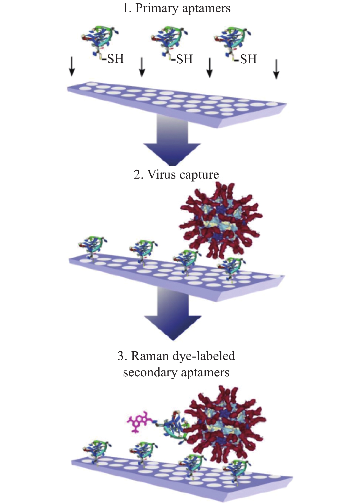
CHEN H, PARK S G, CHOI N, et al. SERS imaging-based aptasensor for ultrasensitive and reproducible detection of influenza virus A[J]. Biosensors and Bioelectronics, 2020, 167: 112496. doi: 10.1016/j.bios.2020.112496
|
| [77] |
CHAUHAN N, SAXENA K, TIKADAR M, et al. Recent advances in the design of biosensors based on novel nanomaterials: an insight[J]. Nanotechnology and Precision Engineering, 2021, 4(4): 045003. doi: 10.1063/10.0006524
|
| [78] |
NGUYEN H A, JUPIN I, DECORSE P, et al. Assembly of gold nanoparticles using turnip yellow mosaic virus as an in-solution SERS sensor[J]. RSC Advances, 2019, 9(55): 32296-32307. doi: 10.1039/C9RA08015E
|
| [79] |
KUKUSHKIN V I, IVANOV N M, NOVOSELTSEVA A A, et al. Highly sensitive detection of influenza virus with SERS aptasensor[J]. PLoS One, 2019, 14(4): e0216247. doi: 10.1371/journal.pone.0216247
|
| [80] |
PENG Y S, LIN C L, LONG L, et al. Charge-transfer resonance and electromagnetic enhancement synergistically enabling MXenes with excellent SERS sensitivity for SARS-CoV-2 S protein detection[J]. Nano-Micro Letters, 2021, 13: 52. doi: 10.1007/s40820-020-00565-4
|
| [81] |
ANTOINE D, MOHAMMADI M, VITT M, et al. Rapid, point-of-care scFv-SERS assay for femtogram level detection of SARS-CoV-2[J]. ACS Sensors, 2022, 7(3): 866-873. doi: 10.1021/acssensors.1c02664
|
| [82] |
LIU B, ZHENG SH Y, LI H T, et al. Ultrasensitive and facile detection of multiple trace antibiotics with magnetic nanoparticles and core-shell nanostar SERS nanotags[J]. Talanta, 2022, 237: 122955. doi: 10.1016/j.talanta.2021.122955
|
| [83] |
SHIN H, OH S, KANG D, et al. Protein quantification and imaging by surface-enhanced Raman spectroscopy and similarity analysis[J]. Advanced Science, 2020, 7(11): 1903638. doi: 10.1002/advs.201903638
|
| [84] |
ZHOU W, TIAN Y F, YIN B CH, et al. Simultaneous surface-enhanced Raman spectroscopy detection of multiplexed microRNA biomarkers[J]. Analytical Chemistry, 2017, 89(11): 6120-6128. doi: 10.1021/acs.analchem.7b00902
|
| [85] |
PANG Y F, WANG CH G, LU L CH, et al. Dual-SERS biosensor for one-step detection of microRNAs in exosome and residual plasma of blood samples for diagnosing pancreatic cancer[J]. Biosensors and Bioelectronics, 2019, 130: 204-213. doi: 10.1016/j.bios.2019.01.039
|
| [86] |
NING C F, WANG L Y, TIAN Y F, et al. Multiple and sensitive SERS detection of cancer-related exosomes based on gold–silver bimetallic nanotrepangs[J]. Analyst, 2020, 145(7): 2795-2804. doi: 10.1039/C9AN02180A
|
| [87] |
LI L, LIU CH, CAO X W, et al. Multiplexing determination of cancer-associated biomarkers by surface-enhanced Raman scattering using ordered gold nanohoneycomb arrays[J]. Bioanalysis, 2017, 9(20): 1561-1572. doi: 10.4155/bio-2016-0237
|
| [88] |
ATANASOV A G, ZOTCHEV S B, DIRSCH V M, et al. Natural products in drug discovery: advances and opportunities[J]. Nature Reviews Drug Discovery, 2021, 20(3): 200-216. doi: 10.1038/s41573-020-00114-z
|
| [89] |
CARTER L J, GARNER L V, SMOOT J W, et al. Assay techniques and test development for COVID-19 diagnosis[J]. ACS Central Science, 2020, 6(5): 591-605. doi: 10.1021/acscentsci.0c00501
|
| [90] |
DE PUIG H, LEE R A, NAJJAR D, et al. Minimally instrumented SHERLOCK (miSHERLOCK) for CRISPR-based point-of-care diagnosis of SARS-CoV-2 and emerging variants[J]. Science Advances, 2021, 7(32): eabh2944. doi: 10.1126/sciadv.abh2944
|
| [91] |
MYHRVOLD C, FREIJE C A, GOOTENBERG J S, et al. Field-deployable viral diagnostics using CRISPR-Cas13[J]. Science, 2018, 360(6387): 444-448. doi: 10.1126/science.aas8836
|
| [92] |
GOOTENBERG J S, ABUDAYYEH O O, KELLNER M J, et al. Multiplexed and portable nucleic acid detection platform with Cas13, Cas12a, and Csm6[J]. Science, 2018, 360(6387): 439-444. doi: 10.1126/science.aaq0179
|
| [93] |
MAKAROVA K S, WOLF Y I, IRANZO J, et al. Evolutionary classification of CRISPR-Cas systems: a burst of class 2 and derived variants[J]. Nature Reviews Microbiology, 2020, 18(2): 67-83. doi: 10.1038/s41579-019-0299-x
|
Figures(9) / Tables(1)


 DownLoad: