| Citation: | WU Zhi-sheng, ZOU Hong-bo, ZHU Wen-wu, QI Wei-ming, WANG Li-qiang, YUAN Bo, YANG Qing, XU Xiao-rong, YAN Hui-hui. Lipid segmentation method based on magnification endoscopy with narrow-band imaging[J]. Chinese Optics, 2024, 17(4): 982-994. doi: 10.37188/CO.EN-2023-0024

|
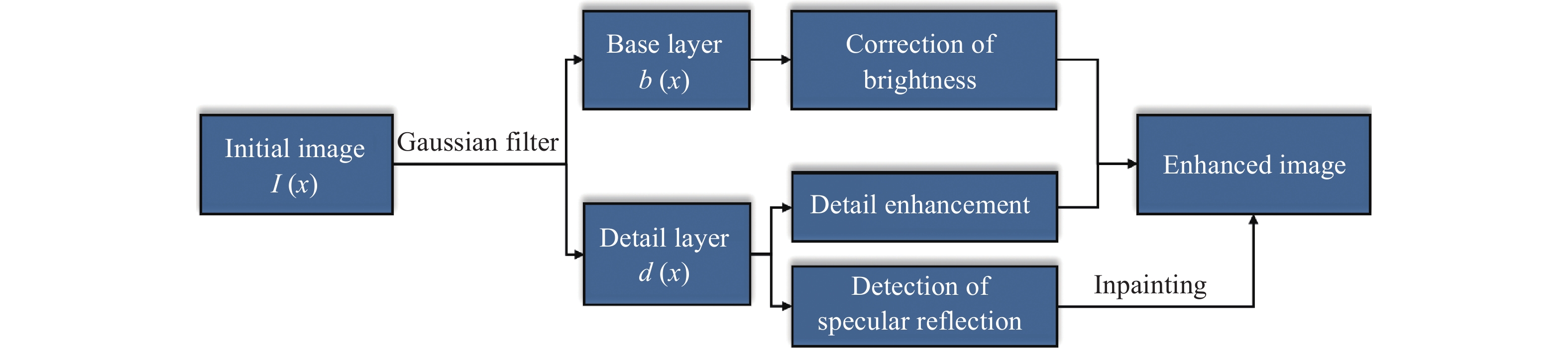
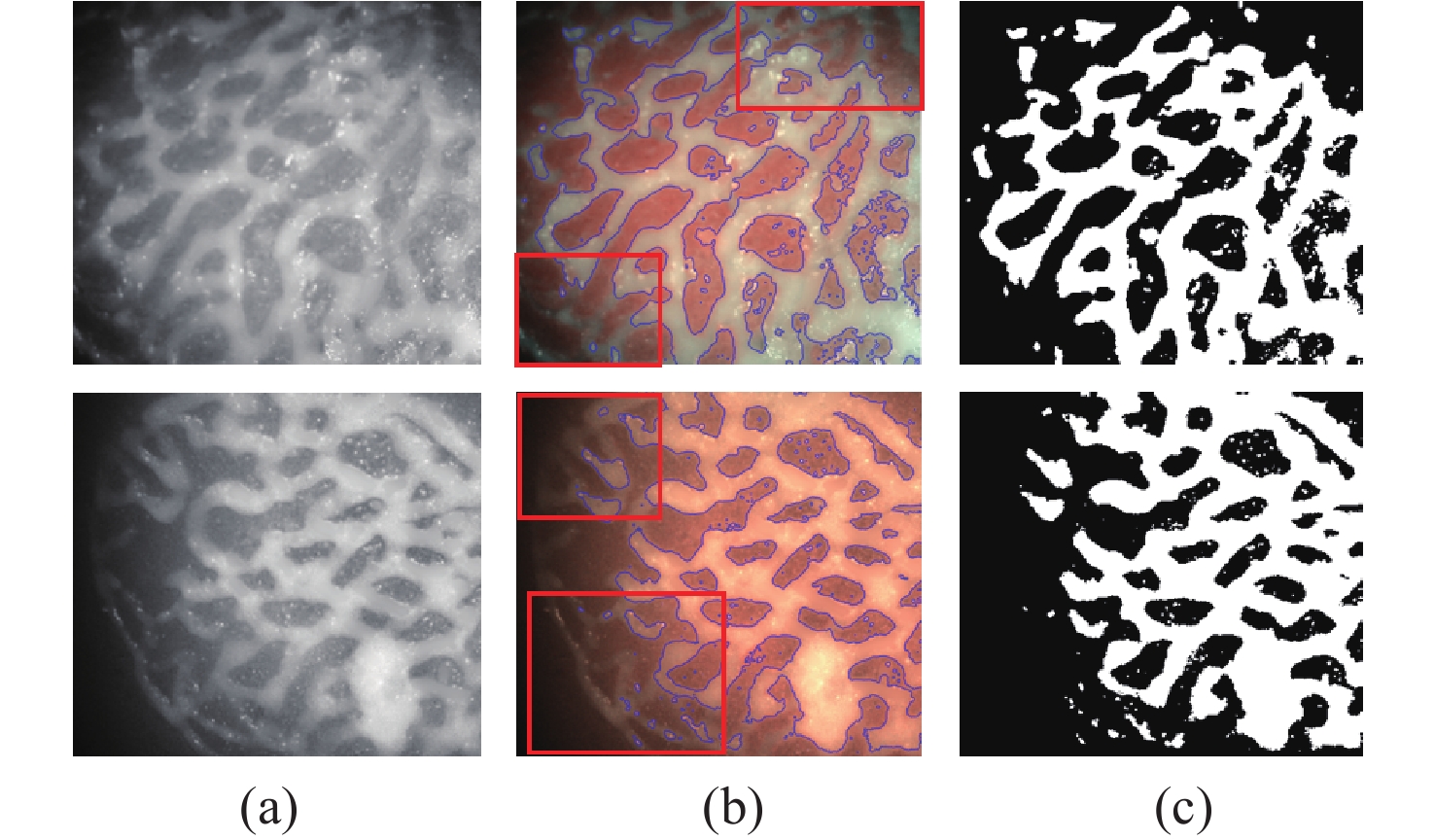
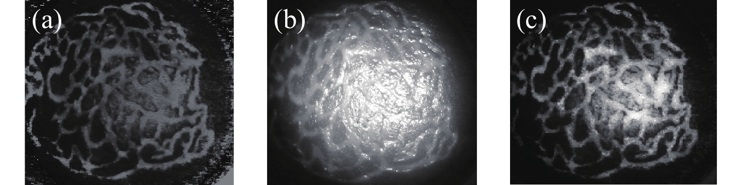
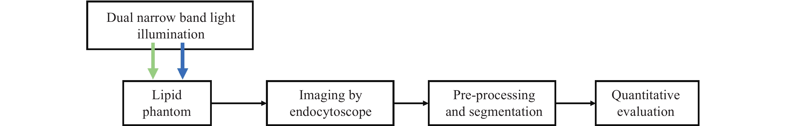
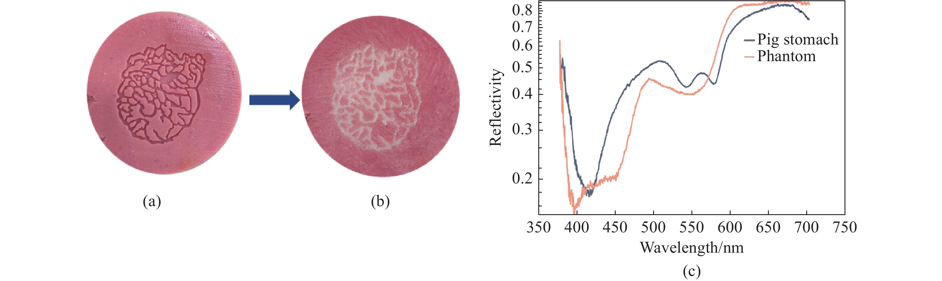
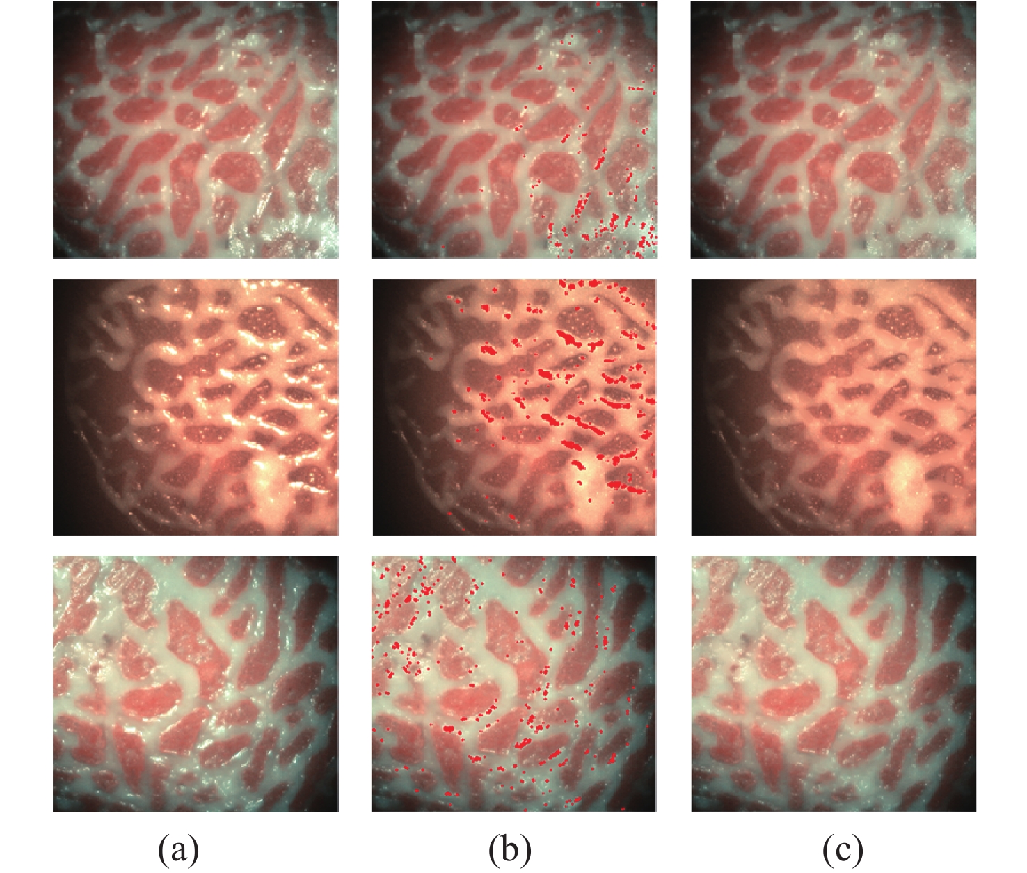
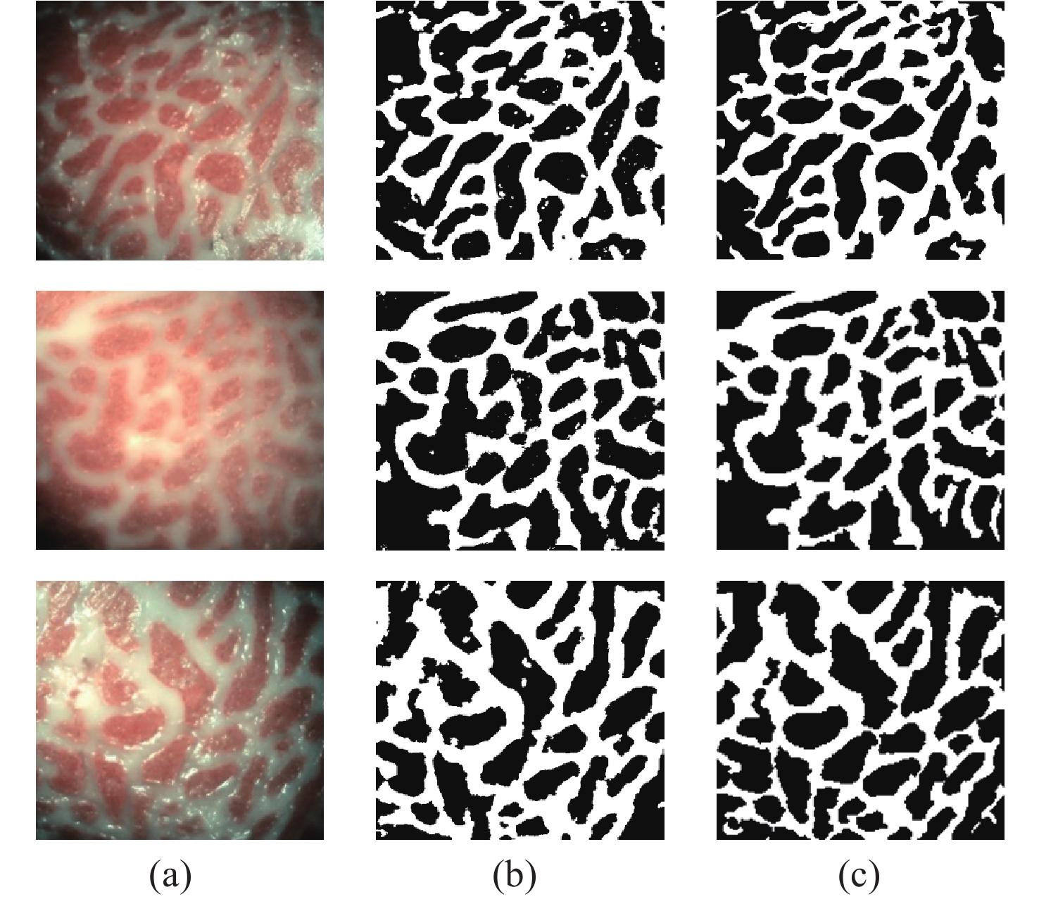
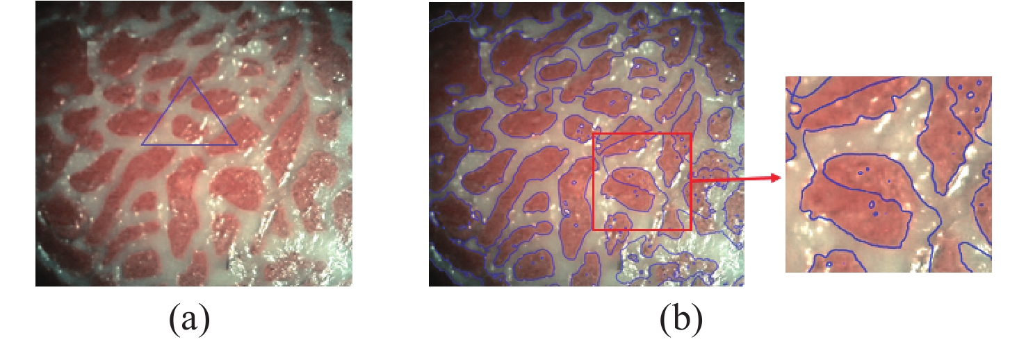
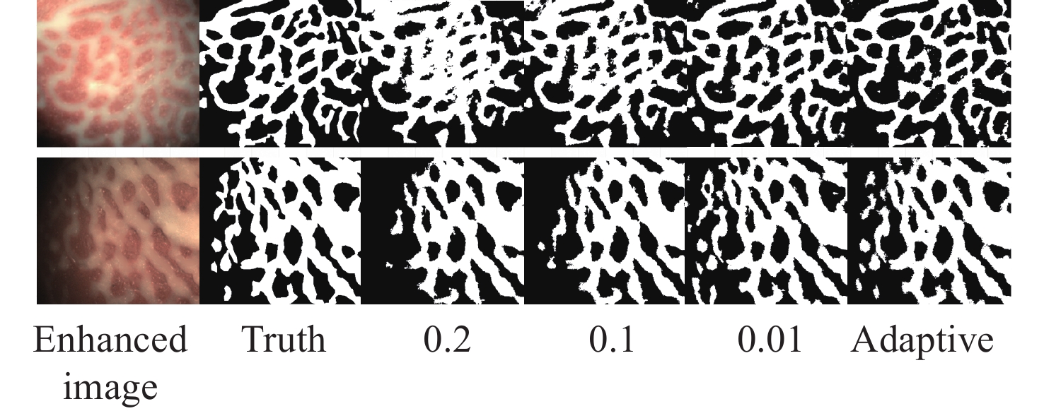
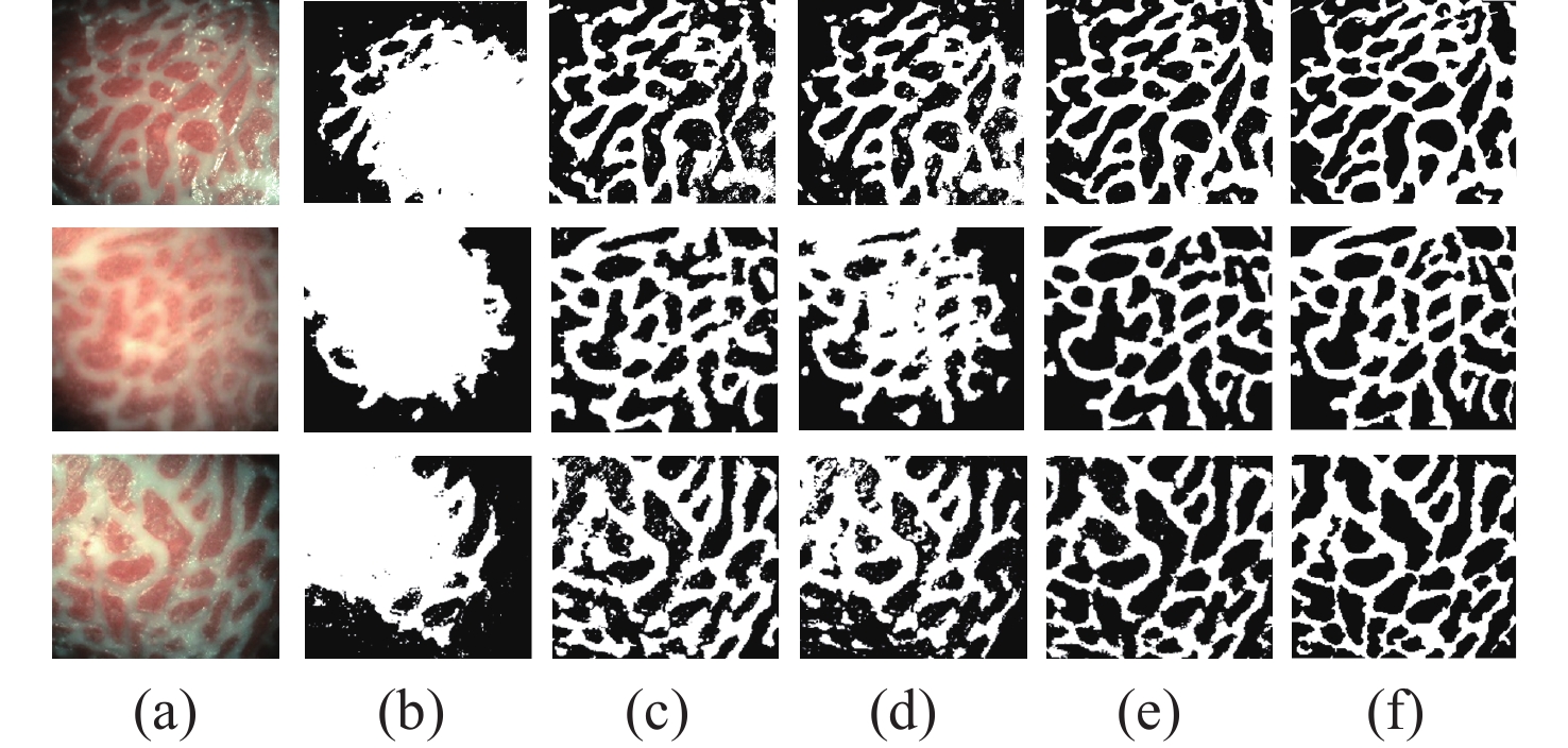
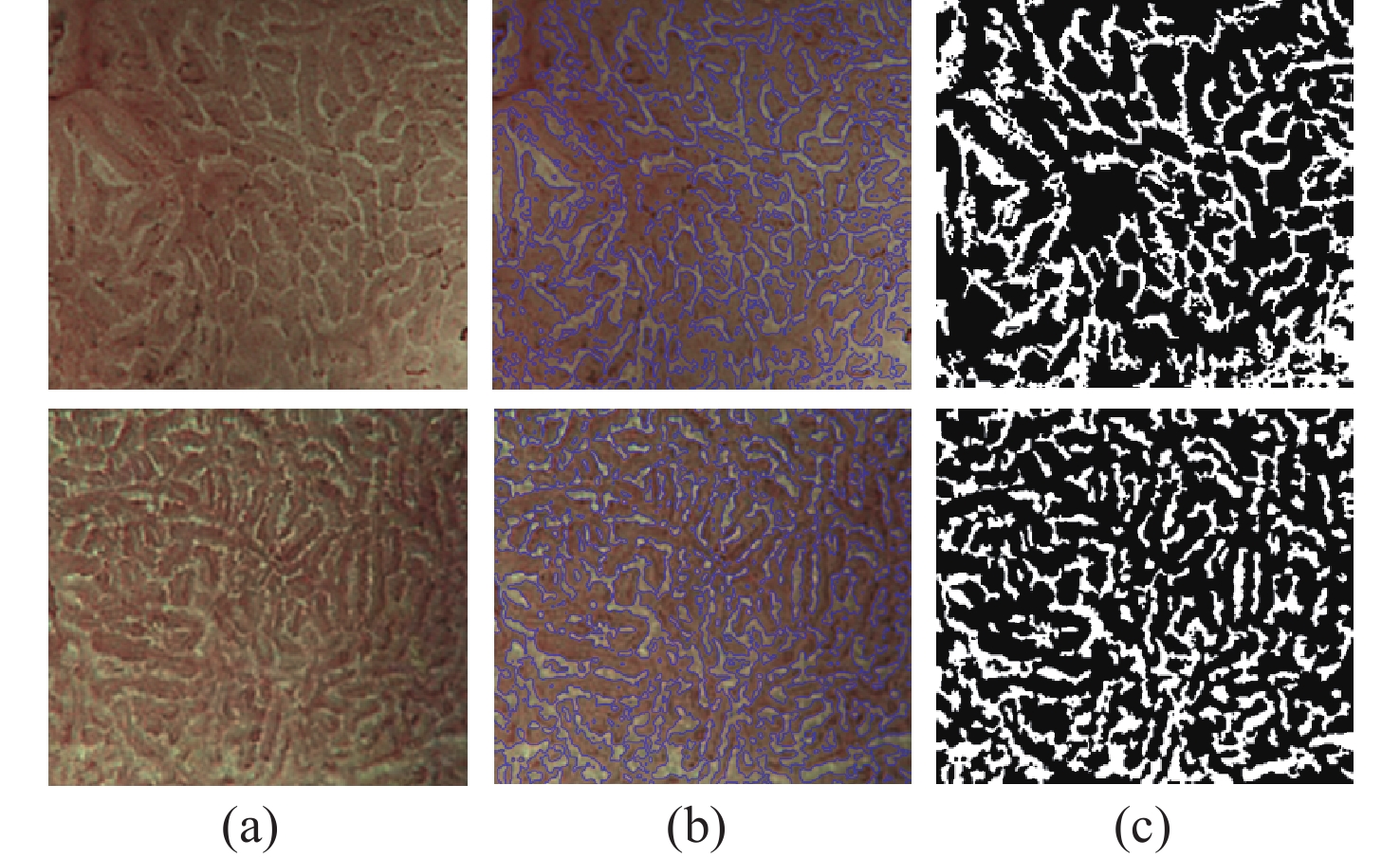
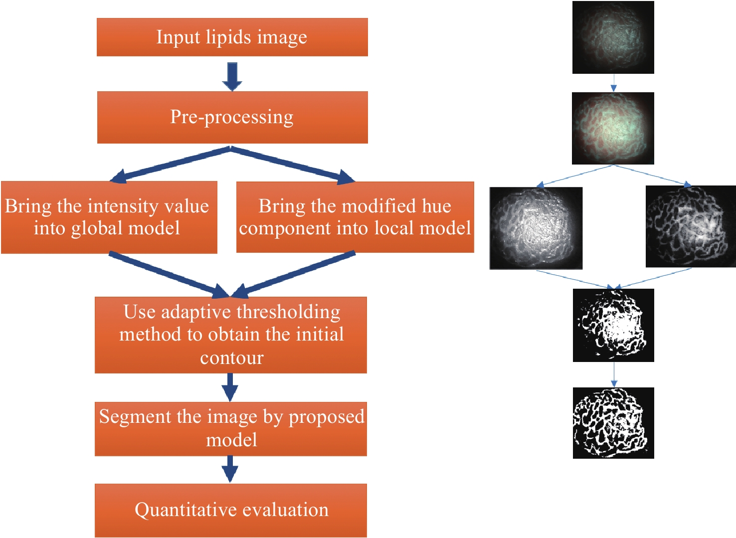
Magnification endoscopy with narrow-band imaging (ME-NBI) has been widely used for cancer diagnosis. However, some microstructures are rendered invisible by a white opaque substance (WOS) composed mainly of lipids. In such lesions, the morphological structure of lipids becomes another marker of tumor grade. We propose a lipid segmentation method. First, the lipid image enhancement algorithm and the specular reflection correction algorithm are introduced. Then, in the framework of the active contour model, the proposed segmentation method extracts local information from modified hue value and global information from intensity value and adaptively obtains the weight factor to segment the lipid region based on the initial contour. This method’s effectiveness is verified by a phantom experiment, which shows that it attained higher than 90% in several key measures: pixel accuracy, sensitivity, and Dice coefficient. The proposed method can accurately reflect the shape of lipids to provide available information for doctors.

| [1] |
SIEGEL R L, MILLER K D, JEMAL A. Cancer statistics, 2020[J]. CA: A Cancer Journal for Clinicians, 2020, 70(1): 7-30. doi: 10.3322/caac.21590
|
| [2] |
MIYAOKA M, YAO K, TANABE H, et al. Diagnosis of early gastric cancer using image enhanced endoscopy: a systematic approach[J]. Translational Gastroenterology and Hepatology, 2020, 5: 50. doi: 10.21037/tgh.2019.12.16
|
| [3] |
KODASHIMA S, FUJISHIRO M, KOIKE K. Image-enhanced endoscopy-NBI, FICE, i-scan[J]. Gastroenterological Endoscopy, 2010, 52(9): 2665-2677.
|
| [4] |
YAMADA S, DOYAMA H, YAO K, et al. An efficient diagnostic strategy for small, depressed early gastric cancer with magnifying narrow-band imaging: a post-hoc analysis of a prospective randomized controlled trial[J]. Gastrointestinal Endoscopy, 2014, 79(1): 55-63. doi: 10.1016/j.gie.2013.07.008
|
| [5] |
ANG T L, FOCK K M, TEO E K, et al. The diagnostic utility of narrow band imaging magnifying endoscopy in clinical practice in a population with intermediate gastric cancer risk[J]. European Journal of Gastroenterology & Hepatology, 2012, 24(4): 362-367.
|
| [6] |
YAO K, ANAGNOSTOPOULOS G K, RAGUNATH K. Magnifying endoscopy for diagnosing and delineating early gastric cancer[J]. Endoscopy, 2009, 41(5): 462-467. doi: 10.1055/s-0029-1214594
|
| [7] |
YAO K, TAKAKI Y, MATSUI T, et al. Clinical application of magnification endoscopy and narrow-band imaging in the upper gastrointestinal tract: new imaging techniques for detecting and characterizing gastrointestinal neoplasia[J]. Gastrointestinal Endoscopy Clinics of North America, 2008, 18(3): 415-433. doi: 10.1016/j.giec.2008.05.011
|
| [8] |
MUTO M, YAO K, KAISE M, et al. Magnifying endoscopy simple diagnostic algorithm for early gastric cancer (MESDA-G)[J]. Digestive Endoscopy, 2016, 28(4): 379-393. doi: 10.1111/den.12638
|
| [9] |
YAO K, IWASHITA A, NAMBU M, et al. Nature of white opaque substance in gastric epithelial neoplasia as visualized by magnifying endoscopy with narrow-band imaging[J]. Digestive Endoscopy, 2012, 24(6): 419-425. doi: 10.1111/j.1443-1661.2012.01314.x
|
| [10] |
YAO K, IWASHITA A, TANABE H, et al. White opaque substance within superficial elevated gastric neoplasia as visualized by magnification endoscopy with narrow-band imaging: a new optical sign for differentiating between adenoma and carcinoma[J]. Gastrointestinal Endoscopy, 2008, 68(3): 574-580. doi: 10.1016/j.gie.2008.04.011
|
| [11] |
NAKAYAMA A, KATO M, TAKATORI Y, et al. How I do it: Endoscopic diagnosis for superficial non-ampullary duodenal epithelial tumors[J]. Digestive Endoscopy, 2020, 32(3): 417-424. doi: 10.1111/den.13538
|
| [12] |
HISABE T, YAO K, IMAMURA K, et al. White opaque substance visualized using magnifying endoscopy with narrow-band imaging in colorectal epithelial neoplasms[J]. Digestive Diseases and Sciences, 2014, 59(10): 2544-2549. doi: 10.1007/s10620-014-3204-5
|
| [13] |
KAWASAKI K, KURAHARA K, YANAI S, et al. Significance of a white opaque substance under magnifying narrow-band imaging colonoscopy for the diagnosis of colorectal epithelial neoplasms[J]. Gastrointestinal Endoscopy, 2015, 82(6): 1097-1104. doi: 10.1016/j.gie.2015.06.023
|
| [14] |
HARA Y, GODA K, HIROOKA S, et al. Association between endoscopic milk-white mucosa, epithelial intracellular lipid droplets, and histological grade of superficial non-ampullary duodenal epithelial tumors[J]. Diagnostics, 2021, 11(5): 769. doi: 10.3390/diagnostics11050769
|
| [15] |
YAMASAKI K, HISABE T, YAO K, et al. White opaque substance, a new optical marker on magnifying endoscopy: usefulness in diagnosing colorectal epithelial neoplasms[J]. Clinical Endoscopy, 2021, 54(4): 570-577. doi: 10.5946/ce.2020.205
|
| [16] |
UEO T, YONEMASU H, YAO K, et al. Histologic differentiation and mucin phenotype in white opaque substance-positive gastric neoplasias[J]. Endoscopy International Open, 2015, 3(6): E597-E604. doi: 10.1055/s-0034-1393177
|
| [17] |
OHTSU K, YAO K, MATSUNAGA K, et al. Lipid is absorbed in the stomach by epithelial neoplasms (adenomas and early cancers): a novel functional endoscopy technique[J]. Endoscopy International Open, 2015, 3(4): E318-E322. doi: 10.1055/s-0034-1392095
|
| [18] |
LIU X Q, WANG CH L, BAI J Y, et al. Hue-texture-embedded region-based model for magnifying endoscopy with narrow-band imaging image segmentation based on visual features[J]. Computer Methods and Programs in Biomedicine, 2017, 145: 53-66. doi: 10.1016/j.cmpb.2017.04.010
|
| [19] |
GANZ M, YANG X Y, SLABAUGH G. Automatic segmentation of polyps in colonoscopic narrow-band imaging data[J]. IEEE Transactions on Biomedical Engineering, 2012, 59(8): 2144-2151. doi: 10.1109/TBME.2012.2195314
|
| [20] |
FIGUEIREDO I N, PINTO L, FIGUEIREDO P N, et al. Unsupervised segmentation of colonic polyps in narrow-band imaging data based on manifold representation of images and Wasserstein distance[J]. Biomedical Signal Processing and Control, 2019, 53: 101577. doi: 10.1016/j.bspc.2019.101577
|
| [21] |
KASS M, WITKIN A, TERZOPOULOS D. Snakes: Active contour models[J]. International Journal of Computer Vision, 1988, 1(4): 321-331. doi: 10.1007/BF00133570
|
| [22] |
BRADLEY D, ROTH G. Adaptive thresholding using the integral image[J]. Journal of Graphics Tools, 2007, 12(2): 13-21. doi: 10.1080/2151237X.2007.10129236
|
| [23] |
WANG L, LI CH M, SUN Q S, et al. Active contours driven by local and global intensity fitting energy with application to brain MR image segmentation[J]. Computerized Medical Imaging and Graphics, 2009, 33(7): 520-531. doi: 10.1016/j.compmedimag.2009.04.010
|
| [24] |
CHAN T F, VESE L A. Active contours without edges[J]. IEEE Transactions on Image Processing, 2001, 10(2): 266-277. doi: 10.1109/83.902291
|
| [25] |
LI CH M, KAO C Y, GORE J C, et al. Implicit active contours driven by local binary fitting energy[C]. 2007 IEEE Conference on Computer Vision and Pattern Recognition, IEEE, 2007: 1-7.
|
| [26] |
JIANG X L, WU X L, XIONG Y, et al. Active contours driven by local and global intensity fitting energies based on local entropy[J]. Optik, 2015, 126(24): 5672-5677. doi: 10.1016/j.ijleo.2015.09.021
|
| [27] |
LI CH M, XU CH Y, GUI CH F, et al. Level set evolution without re-initialization: a new variational formulation[C]. 2005 IEEE Computer Society Conference on Computer Vision and Pattern Recognition (CVPR'05), IEEE, 2005: 430-436.
|
| [28] |
ZHANG L, PENG X G, LI G, et al. A novel active contour model for image segmentation using local and global region-based information[J]. Machine Vision and Applications, 2017, 28(1-2): 75-89. doi: 10.1007/s00138-016-0805-3
|
| [29] |
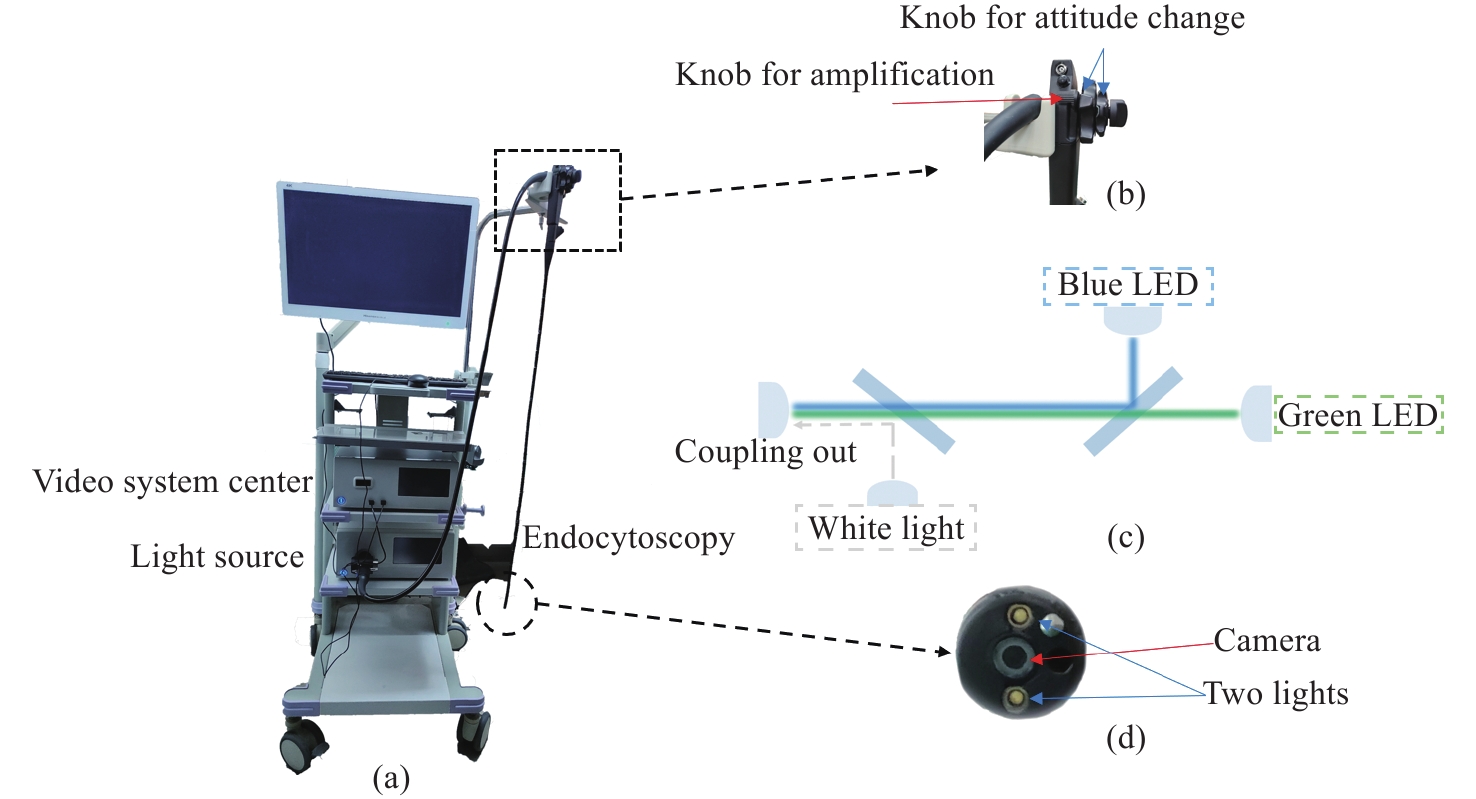
ZHANG W, NIU CH Y, YOU X H, et al. Endocytoscopic imaging system with high magnification and large field of view[J]. Acta Optica Sinica, 2021, 41(17): 1717001. doi: 10.3788/AOS202141.1717001
|
| [30] |
POGUE B W, PATTERSON M S. Review of tissue simulating phantoms for optical spectroscopy, imaging and dosimetry[J]. Journal of Biomedical Optics, 2006, 11(4): 041102. doi: 10.1117/1.2335429
|
| [31] |
RUSSELL B C, TORRALBA A, MURPHY K P, et al. LabelMe: A database and web-based tool for image annotation[J]. International Journal of Computer Vision, 2008, 77(1): 157-173.
|
Figures(14) / Tables(2)


 DownLoad: