


Design of a miniature head-mounted fluorescence microscope based on gradient refractive index lenses
-
摘要:
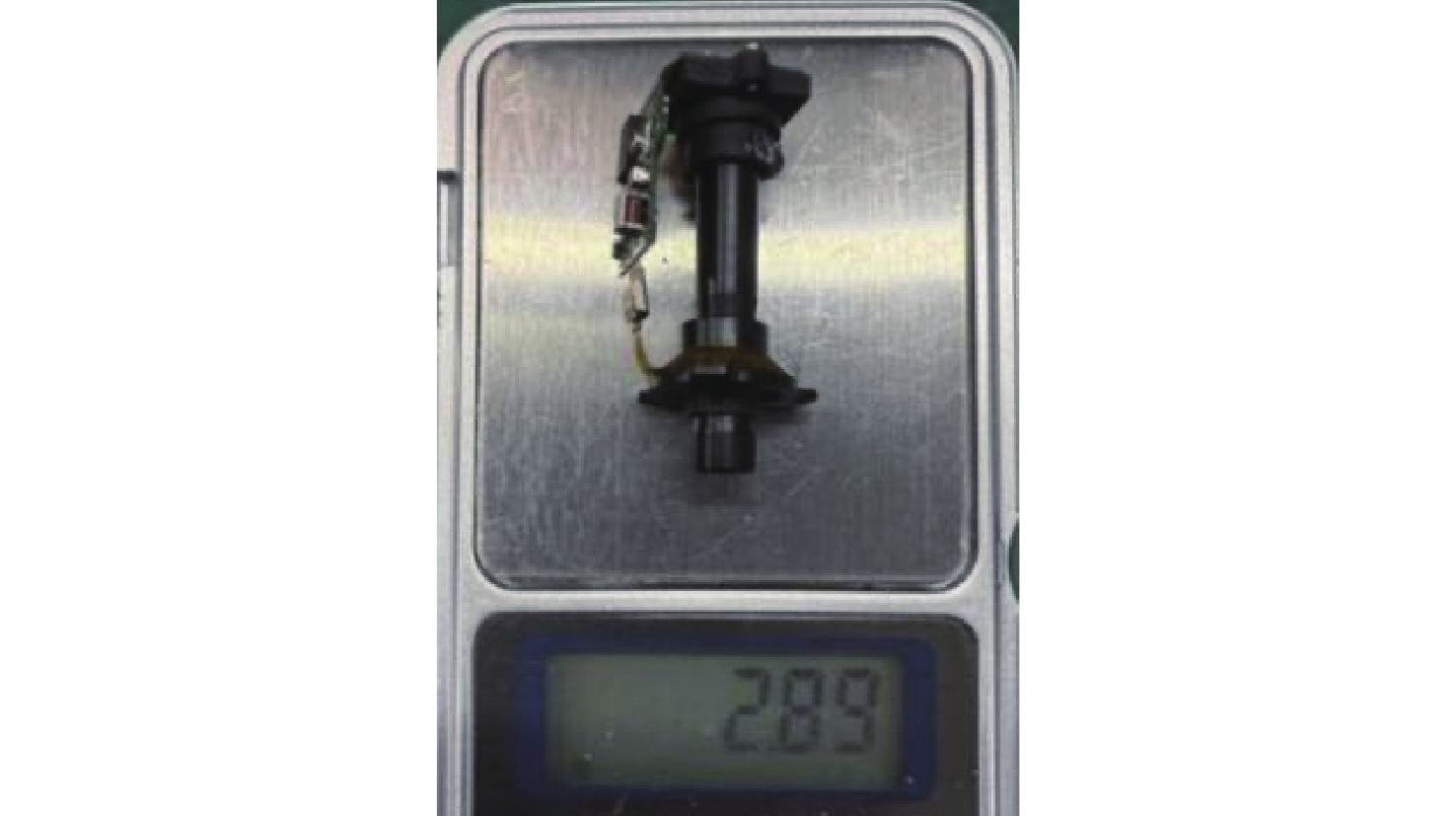
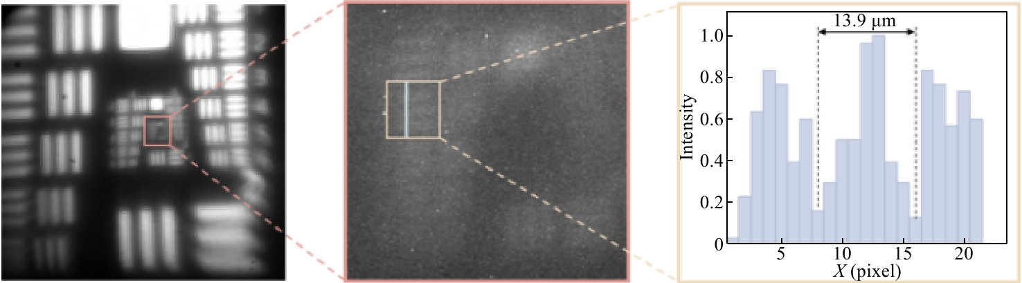
在对自由移动的动物进行脑神经实时观测时,微型头戴式荧光显微镜是当前最为前沿的脑科学观测仪器之一。但是,现今多数微型荧光显微镜的体积和重量受到严格限制,使得视场较小,无法同时观测多个脑区的神经活动,而少数视场大的产品重量较大,无法佩戴在小型动物身上。本研究使用轻量化、平面化和成像质量高的梯度折射率透镜进行大视场微型荧光显微镜设计,在保证大视场的前提下,减小了显微镜的重量。推导了倾斜光线入射梯度折射率透镜时的离轴像差公式,分析了梯度折射率透镜的折射率排布模型和像差校正情况。最终设计的微型荧光显微镜的视场为4 mm×4 mm,NA为0.1,样机重量仅为2.89 g,中心视场分辨率为13.9 μm,初步达到了自由移动小鼠的脑神经细胞分辨率。
Abstract:In real-time brain neural observation of freely moving animals, the miniature head-mounted fluorescence microscope is currently one of the most advanced brain science observation instruments. However, most existing miniature fluorescence microscopes, in order to meet strict size and weight constraints, have a limited field of view, making it impossible to simultaneously observe neural activity in multiple brain regions. On the other hand, a few products with a larger field of view are too heavy to be worn on small animals. This study employs lightweight, planar, and high-quality gradient refractive index lenses to reduce the microscope's weight while ensuring a large field of view. Using gradient refractive index lenses for the design of a large-field-of-view miniature fluorescence microscope, this research derives the off-axis aberration formula for oblique light incidence on gradient refractive index lenses, analyzes the refractive index distribution model and aberration correction of these lenses, and designs a miniature fluorescence microscope with a 4 mm×4 mm field of view, a numerical aperture (NA) of 0.1, and a prototype weight of only 2.89 g. The central visual field resolution is 13.9 μm, preliminarily achieving the resolution for neural cells in freely moving mice.
-
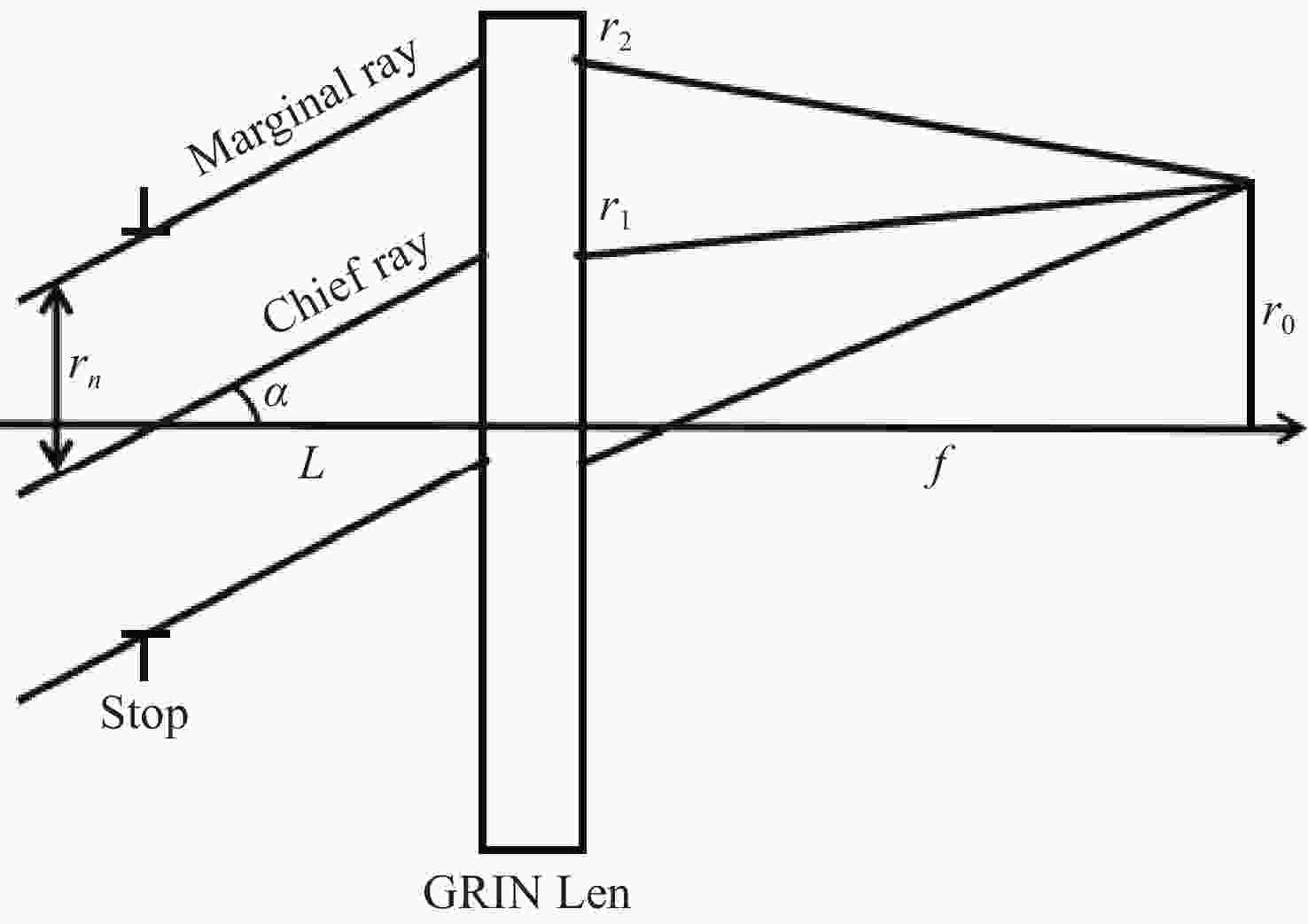
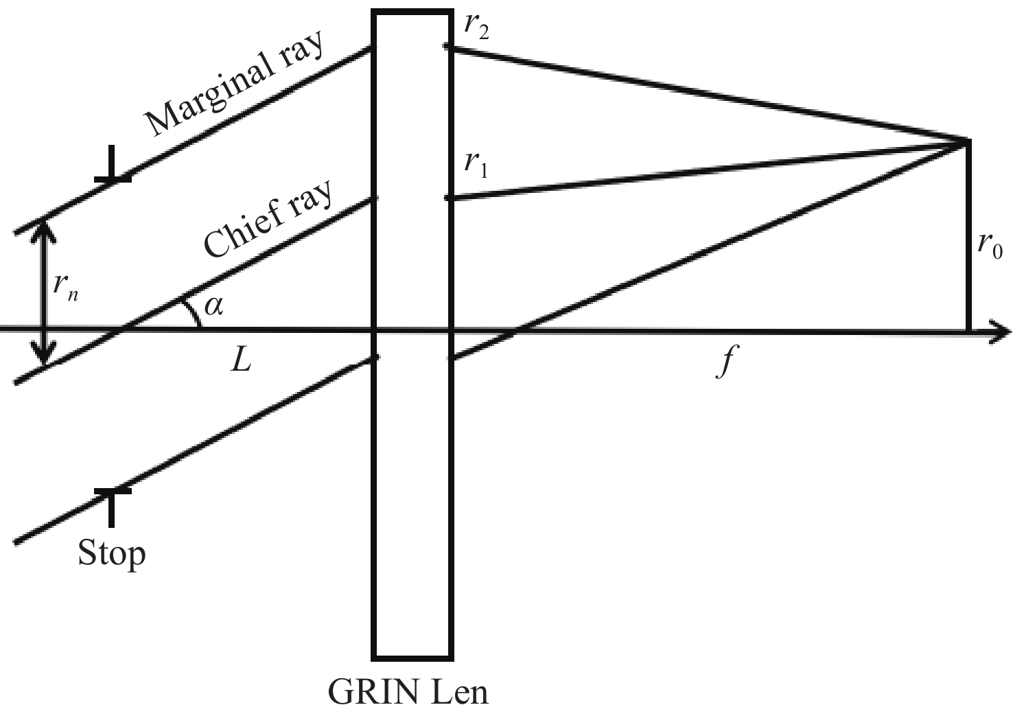
图 1 倾斜光线经过梯度折射率透镜的模拟光路
Figure 1. Simulated optical path of oblique light rays through a gradient refractive index lens
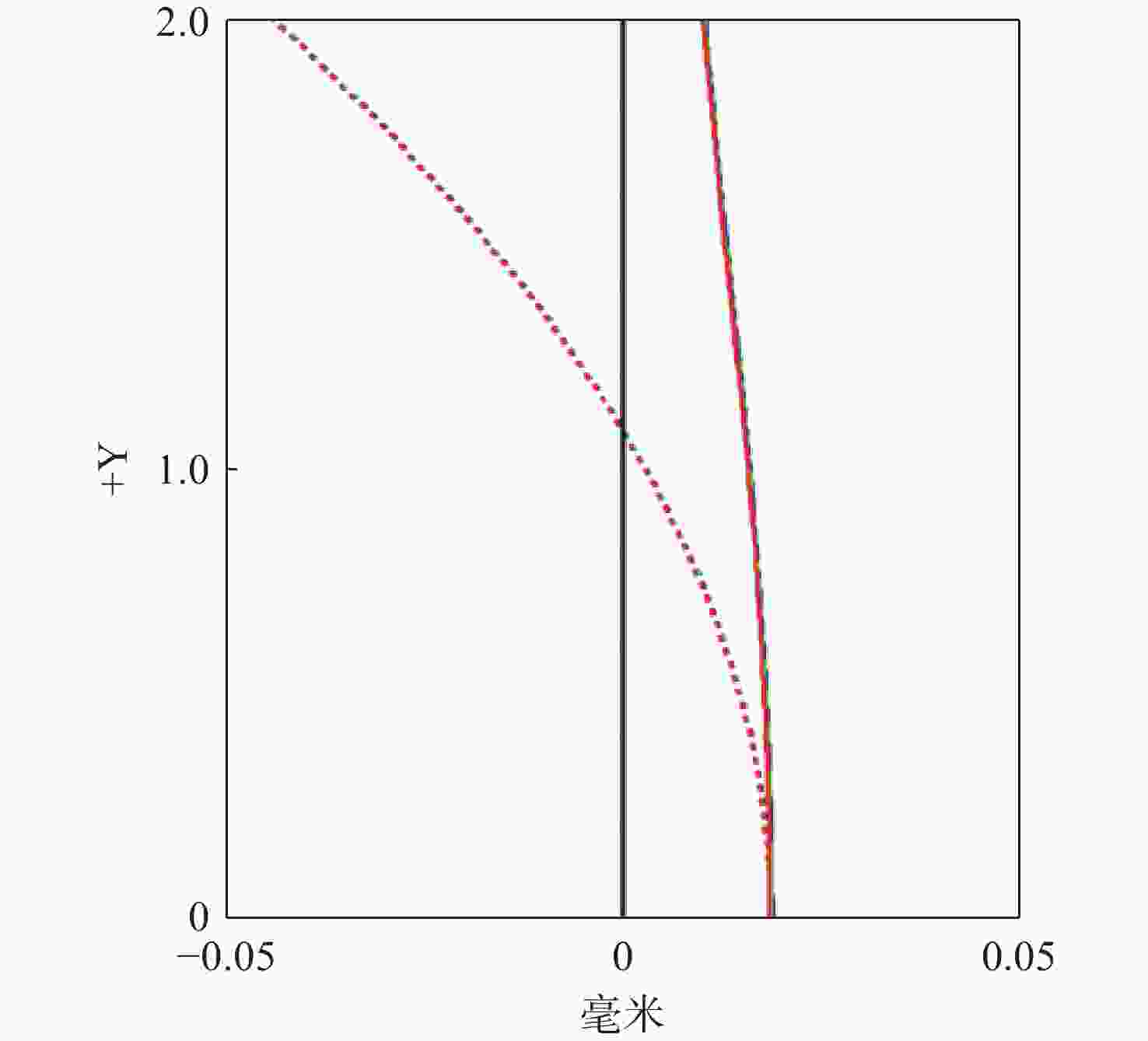
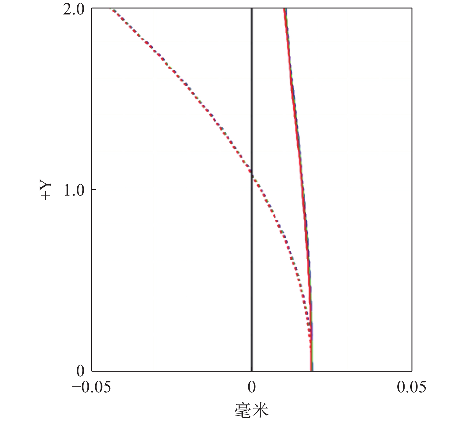
图 6 显微镜的场曲和像散曲线
Figure 6. Field curvature and astigmatism curves of the miniature fluorescence microscope
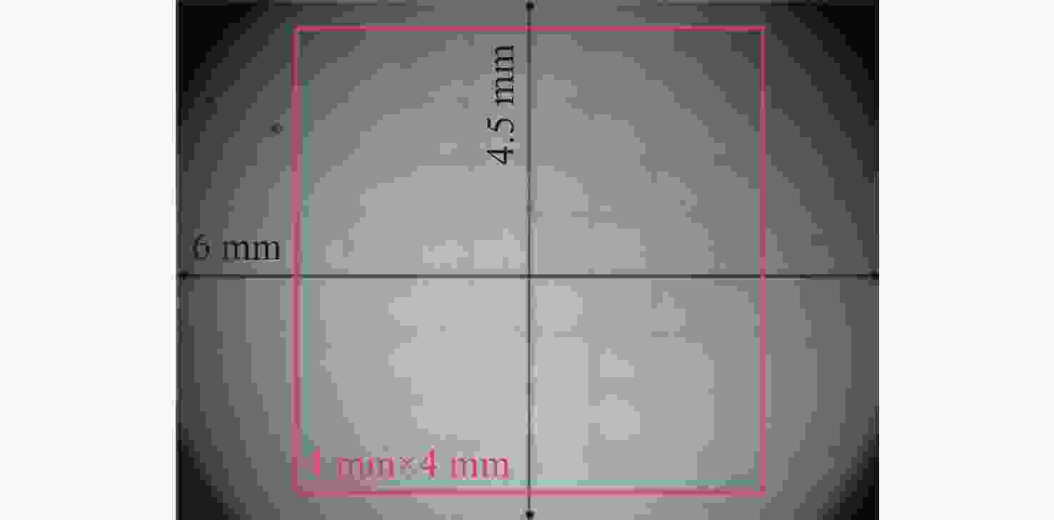
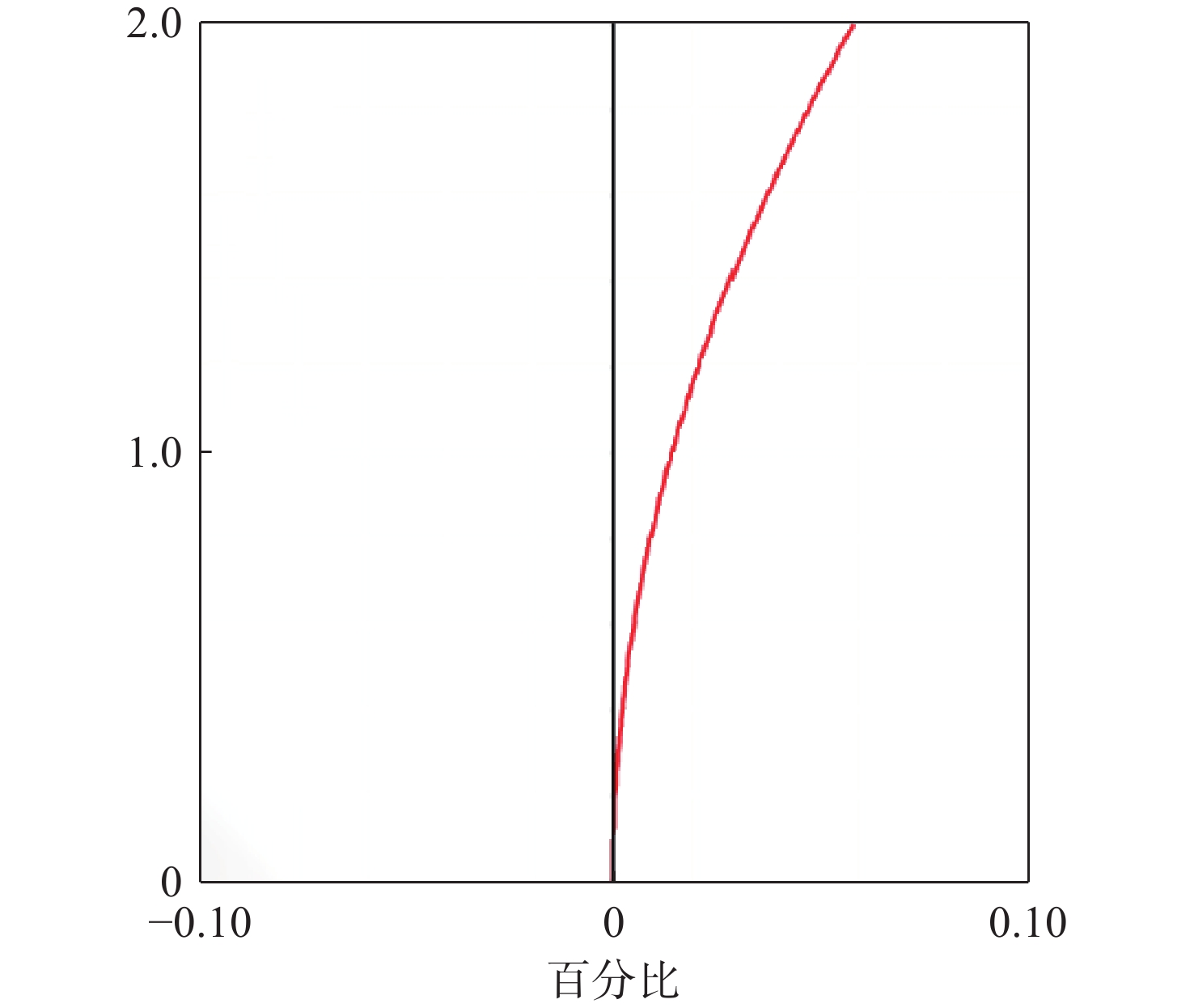
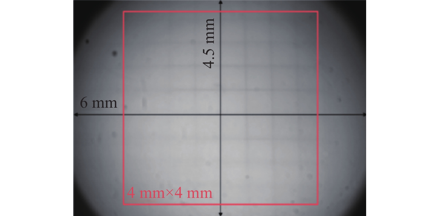
图 11 畸变测试结果
Figure 11. Distortion test results of the miniature fluorescence microscope
-
[1] 刘晓宇, 刘紫千, 斯科, 等. 微型化显微成像系统的关键技术及研究进展(特邀)[J]. 激光与光电子学进展, 2024, 61(2): 0211009. doi: 10.3788/LOP232709LIU X Y, LIU Z Q, SI K, et al. Key technologies and progresses of miniaturized microscopic imaging system (invited)[J]. Laser & Optoelectronics Progress, 2024, 61(2): 0211009. (in Chinese). doi: 10.3788/LOP232709 [2] KAMATH V, BHAT V G, RAJU G, et al. Application of fluorescence lifetime imaging-integrated deep learning analysis for cancer research[J]. Light: Advanced Manufacturing, 2025, 6(3): 49. doi: 10.37188/lam.2025.049 [3] YU H, SENARATHNA J, TYLER B M, et al. Miniaturized optical neuroimaging in unrestrained animals[J]. NeuroImage, 2015, 113: 397-406. doi: 10.1016/j.neuroimage.2015.02.070 [4] CHEN SH Y, WANG Z CH, ZHANG D, et al. Miniature fluorescence microscopy for imaging brain activity in freely-behaving animals[J]. Neuroscience Bulletin, 2020, 36(10): 1182-1190. doi: 10.1007/s12264-020-00561-z [5] HOU Y W, WANG W Y, FU Y ZH, et al. Multi-resolution analysis enables fidelity-ensured deconvolution for fluorescence microscopy[J]. eLight, 2024, 4(1): 14. doi: 10.1186/s43593-024-00073-7 [6] YANNY K, ANTIPA N, LIBERTI W, et al. Miniscope3D: optimized single-shot miniature 3D fluorescence microscopy[J]. Light: Science & Applications, 2020, 9(1): 171. [7] 胡鹏涛, 高若谦, 葛明锋, 等. 流动相单分子免疫检测系统的设计[J]. 中国光学(中英文), 2025, 18(5): 1055-1065. doi: 10.37188/CO.2025-0045HU P T, GAO R Q, GE M F, et al. Design of flow-phase single-molecule immunoassay detection system[J]. Chinese Optics, 2025, 18(5): 1055-1065. (in Chinese). doi: 10.37188/CO.2025-0045 [8] 王浩然, 常雪莹, 陈启龙, 等. 用于集成成像3D显示的模式控制型液晶微透镜阵列[J]. 液晶与显示, 2025, 40(8): 1132-1144.WANG H R, CHANG X Y, CHEN Q L, et al. Liquid crystal micro lens array with modal control for integrated imaging 3D display[J]. Chinese Journal of Liquid Crystals and Displays, 2025, 40(8): 1132-1144. [9] 付强, 张智淼, 赵尚男, 等. 微型头戴式单光子荧光显微成像技术研究进展[J]. 中国光学 (中英文), 2023, 16(5): 1010-1021.FU Q, ZHANG ZH M, ZHAO SH N, et al. Research progress of miniature head-mounted single photon fluorescence microscopic imaging technique[J]. Chinese Optics, 2023, 16(5): 1010-1021. (in Chinese). [10] RYNES M L, SURINACH D A, LINN S, et al. Miniaturized head-mounted microscope for whole-cortex mesoscale imaging in freely behaving mice[J]. Nature Methods, 2021, 18(4): 417-425. [11] PARK D H, JOO B C, CHOI K R, et al. 3D printed miniature attenuated total internal reflection assisted fluorescence microscopy[J]. Sci Rep, 2025, 15: 7683. [12] EROFEV A I, VINOKUROV E K, ANTIFEEV I E, et al. Integration of single-photon miniature fluorescence microscopy and electrophysiological recording methods for in vivo studying hippocampal neuronal activity[J]. J Evol Biochem Phys, 2024, 60: 1586-1606. [13] GERASIMOV E, PCHITSKAYA E, VLASOVA O, et al. Dynamic changes in the hippocampal neuronal circuits activity following acute stress revealed by miniature fluorescence microscopy imaging[J]. Mol Brain, 2024, 17: 92. doi: 10.1038/s41598-025-92204-4 [14] GERASIMOV E, MITENEV A, PCHITSKAYA E, et al. NeuroActivityToolkit—Toolbox for Quantitative Analysis of Miniature Fluorescent Microscopy Data[J]. Journal of Imaging, 2023, 9(11): 243. doi: 10.1134/S0022093024040264 [15] ZHAO P P, GUO C L, XIE M, et al. An open-source, large field-of-view epifluorescence miniature microscope for mice capable of single-cell resolution and multi-brain region imaging[J]. bioRxiv: 2024.08.16.608328. [16] 张智淼, 王承邈, 谢冕, 等. 基于超构透镜的微型头戴式荧光显微镜设计[J]. 中国光学(中英文), 2024, 17(03): 512-520. doi: 10.3390/jimaging9110243ZHANG ZH M, WANG CH M, XIE M, et al. Design of miniature head-mounted fluorescence microscope based on metalens[J]. Chinese Optics, 2024, 17(3): 512-520. (in Chinese). doi: 10.3390/jimaging9110243 [17] SCOTT B B, THIBERGE S Y, GUO C Y, et al. Imaging cortical dynamics in GCaMP transgenic rats with a headmounted widefield macroscope[J]. Neuron, 2018, 100(5): 1045-1058. e5. [18] SUPEKAR O D, SIAS A, HANSEN S R, et al. Miniature structured illumination microscope for in vivo 3D imaging of brain structures with optical sectioning[J]. Biomedical Optics Express, 2022, 13(4): 2530-2541. [19] GUO CH L, BLAIR G J, SEHGAL M, et al. Miniscope-LFOV: a large-field-of-view, single-cell-resolution, miniature microscope for wired and wire-free imaging of neural dynamics in freely behaving animals[J]. Science Advances, 2023, 9(16): eadf3918. [20] BAGRAMYAN A. Lightweight 1-photon miniscope for imaging in freely behaving animals at subcellular resolution[J]. IEEE Photonics Technology Letters, 2020, 32(15): 909-912. [21] WANG Y ZH, MA ZH T, LI W ZH, et al. Cable-free brain imaging with miniature wireless microscopes[J]. Journal of Biomedical Optics, 2023, 28(2): 026503. [22] XUE F, Li F, Zhang K M, et al. Multi-region calcium imaging in freely behaving mice with ultra-compact head-mounted fluorescence microscopes[J]. National Science Review, 2024, 11(1): nwad286. doi: 10.1126/sciadv.adg3918 [23] YANG T. Freeform gradient-index optics with applications in rotationally variant systems[D]. Rochester: University of Rochester, 2018. doi: 10.1109/LPT.2020.3004283 [24] YOUNG M. Zone plates and their aberrations[J]. Journal of the Optical Society of America, 1972, 62(8): 972-976. [25] GROSS H. Handbook of Optical Systems (Volume 3: Aberration Theory and Correction of Optical Systems)[M]. Weinheim: John Wiley & Sons Inc, 2007. doi: 10.1117/1.jbo.28.2.026503 -

 下载:


计量
- 文章访问数: 238
- HTML全文浏览量: 124
- PDF下载量: 9
- 被引次数: 0
