| Citation: | ZHOU Ri, WANG Chen-guang, LU Ge-yu. Advances in organic fluorescent probes for super-resolution imaging of cellular lipid droplets[J]. Chinese Optics, 2022, 15(6): 1228-1242. doi: 10.37188/CO.2022-0077

|
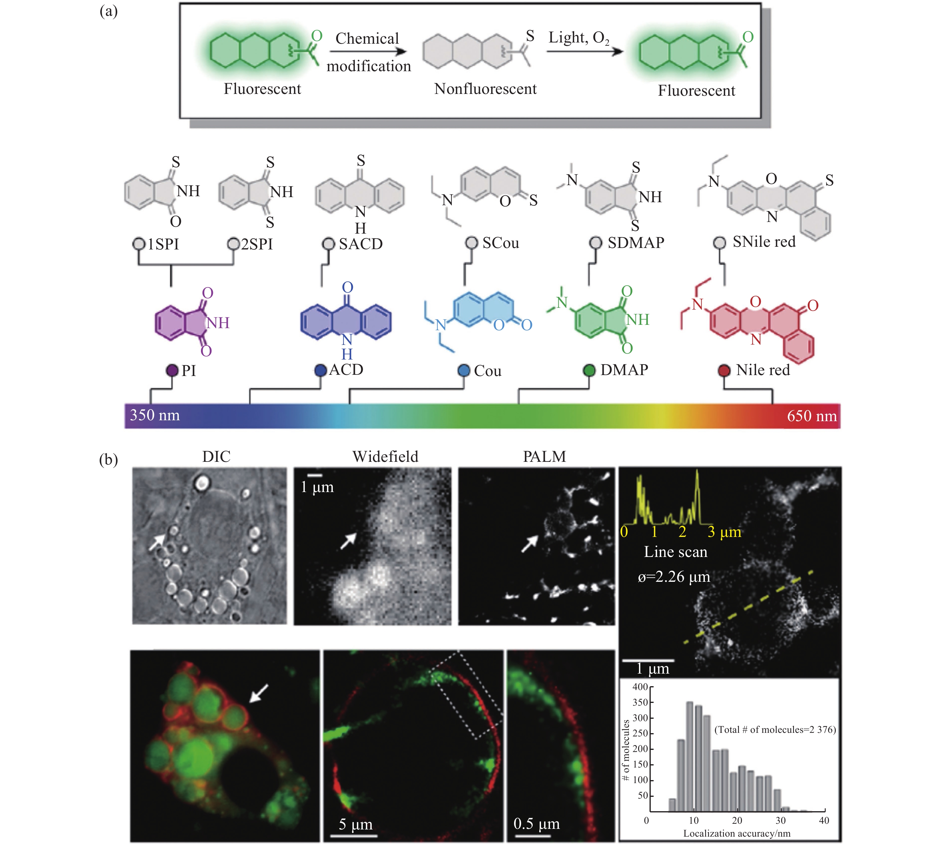
Lipid droplets are a kind of spherical organelle in eukaryotic cells and are relevant to many cellular physiological processes. Fluorescence imaging techniques are one of the most powerful tools to visualize and study lipid droplets. However, conventional wide-field microscopy and confocal microscopy can only provide a resolution of about 250 nm due to the limitation of optical diffraction. This resolution is quite insufficient for visualizing the small lipid droplets, especially the nascent ones (size of about 30~60 nm). Emerging super-resolution microscopes that can break the diffraction limit (such as stimulated emission depletion microscopy, structured illumination microscopy and photoactivated localization microscopy) have gradually attracted much interest in recent years. To obtain high-resolution fluorescence images of lipid droplets, the advanced fluorescent probes which meet the special requirements of the corresponding super-resolution microscopes are highly essential. This review paper will briefly introduce the working principles of various super-resolution microscopes, discuss the special requirements on the photophysical properties of fluorescent probes, and systematically summarize the research progress of super-resolution imaging of lipid droplets by employing these fluorescent probes. Meanwhile, this review will compare the advantages and shortcomings of different super-resolution techniques for lipid droplets imaging, and prospect their future possible trends.

| [1] |
OLZMANN J A, CARVALHO P. Dynamics and functions of lipid droplets[J]. Nature Reviews Molecular Cell Biology, 2019, 20(3): 137-155. doi: 10.1038/s41580-018-0085-z
|
| [2] |
THIAM A R, BELLER M. The why, when and how of lipid droplet diversity[J]. Journal of Cell Science, 2017, 130(2): 315-324.
|
| [3] |
CHOUDHARY V, OJHA N, GOLDEN A, et al. A conserved family of proteins facilitates nascent lipid droplet budding from the ER[J]. Journal of Cell Biology, 2015, 211(2): 261-271. doi: 10.1083/jcb.201505067
|
| [4] |
JACQUIER N, CHOUDHARY V, MARI M, et al. Lipid droplets are functionally connected to the endoplasmic reticulum in Saccharomyces cerevisiae[J]. Journal of Cell Science, 2011, 124(14): 2424-2437. doi: 10.1242/jcs.076836
|
| [5] |
KASSAN A, HERMS A, FERNÁNDEZ-VIDAL A, et al. Acyl-CoA synthetase 3 promotes lipid droplet biogenesis in ER microdomains[J]. Journal of Cell Biology, 2013, 203(6): 985-1001. doi: 10.1083/jcb.201305142
|
| [6] |
BUHMAN K K, CHEN H C, FARESE R V JR. The enzymes of neutral lipid synthesis[J]. Journal of Biological Chemistry, 2001, 276(44): 40369-40372. doi: 10.1074/jbc.R100050200
|
| [7] |
THIAM A R, FARESE R V JR, WALTHER T C. The biophysics and cell biology of lipid droplets[J]. Nature Reviews Molecular Cell Biology, 2013, 14(12): 775-786. doi: 10.1038/nrm3699
|
| [8] |
WALTHER T C, FARESE R V JR. Lipid droplets and cellular lipid metabolism[J]. Annual Review of Biochemistry, 2012, 81: 687-714. doi: 10.1146/annurev-biochem-061009-102430
|
| [9] |
FARESE R V JR, WALTHER T C. Lipid droplets finally get a little R-E-S-P-E-C-T[J]. Cell, 2009, 139(5): 855-860. doi: 10.1016/j.cell.2009.11.005
|
| [10] |
ROITENBERG N, COHEN E. Lipid assemblies at the crossroads of aging, proteostasis, and neurodegeneration[J]. Trends in Cell Biology, 2019, 29(12): 954-963. doi: 10.1016/j.tcb.2019.09.003
|
| [11] |
KRAHMER N, FARESE R V JR, WALTHER T C. Balancing the fat: lipid droplets and human disease[J]. EMBO Molecular Medicine, 2013, 5(7): 973-983. doi: 10.1002/emmm.201100671
|
| [12] |
ONAL G, KUTLU O, GOZUACIK D, et al. Lipid droplets in health and disease[J]. Lipids in Health and Disease, 2017, 16(1): 128. doi: 10.1186/s12944-017-0521-7
|
| [13] |
LIU Q P, LUO Q, HALIM A, et al. Targeting lipid metabolism of cancer cells: a promising therapeutic strategy for cancer[J]. Cancer Letters, 2017, 401: 39-45. doi: 10.1016/j.canlet.2017.05.002
|
| [14] |
COLLOT M, FAM T K, ASHOKKUMAR P, et al. Ultrabright and fluorogenic probes for multicolor imaging and tracking of lipid droplets in cells and tissues[J]. Journal of the American Chemical Society, 2018, 140(16): 5401-5411. doi: 10.1021/jacs.7b12817
|
| [15] |
GUO L F, TIAN M G, ZHANG ZH Y, et al. Simultaneous two-color visualization of lipid droplets and endoplasmic reticulum and their interplay by single fluorescent probes in lambda mode[J]. Journal of the American Chemical Society, 2021, 143(8): 3169-3179. doi: 10.1021/jacs.0c12323
|
| [16] |
SHI L, LI K, LI L L, et al. Novel easily available purine-based AIEgens with colour tunability and applications in lipid droplet imaging[J]. Chemical Science, 2018, 9(48): 8969-8974. doi: 10.1039/C8SC03369B
|
| [17] |
ZHANG CH, LI J J, LAN L, et al. Quantification of lipid metabolism in living cells through the dynamics of lipid droplets measured by stimulated raman scattering imaging[J]. Analytical Chemistry, 2017, 89(8): 4502-4507. doi: 10.1021/acs.analchem.6b04699
|
| [18] |
ZHANG CH, BOPPART S A. Dynamic signatures of lipid droplets as new markers to quantify cellular metabolic changes[J]. Analytical Chemistry, 2020, 92(24): 15943-15952. doi: 10.1021/acs.analchem.0c03366
|
| [19] |
TAKI M, KAJIWARA K, YAMAGUCHI E, et al. Fused thiophene-S, S-dioxide-based super-photostable fluorescent marker for lipid droplets[J]. ACS Materials Letters, 2021, 3(1): 42-49. doi: 10.1021/acsmaterialslett.0c00451
|
| [20] |
XU Y Z, ZHANG H K, ZHANG N, et al. An easily synthesized AIE luminogen for lipid droplet-specific super-resolution imaging and two-photon imaging[J]. Materials Chemistry Frontiers, 2021, 5(4): 1872-1883. doi: 10.1039/D0QM00682C
|
| [21] |
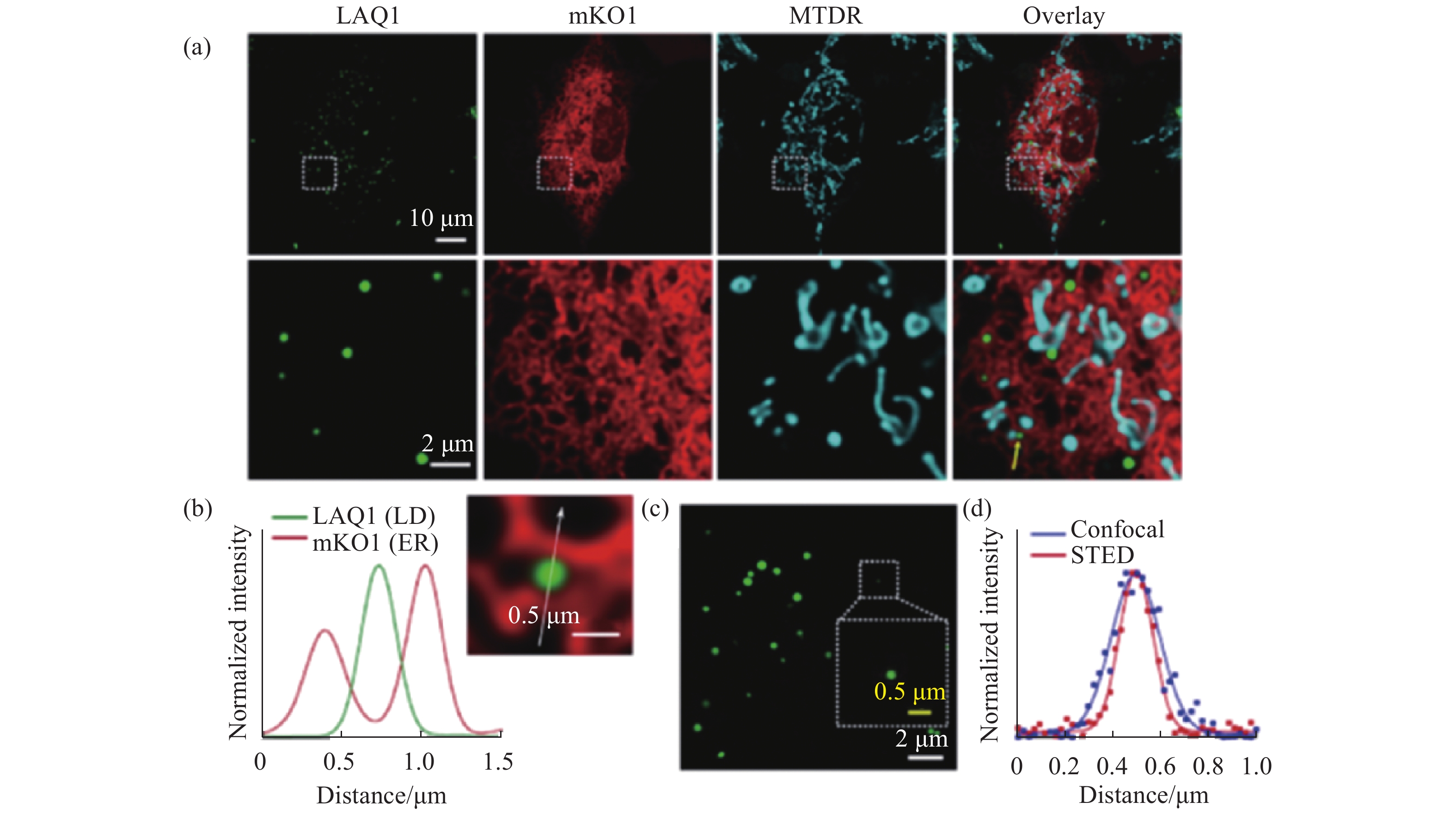
ZHOU R, WANG CH G, LIANG X SH, et al. Stimulated emission depletion (STED) super-resolution imaging with an advanced organic fluorescent probe: visualizing the cellular lipid droplets at the unprecedented nanoscale resolution[J]. ACS Materials Letters, 2021, 3(5): 516-524. doi: 10.1021/acsmaterialslett.1c00143
|
| [22] |
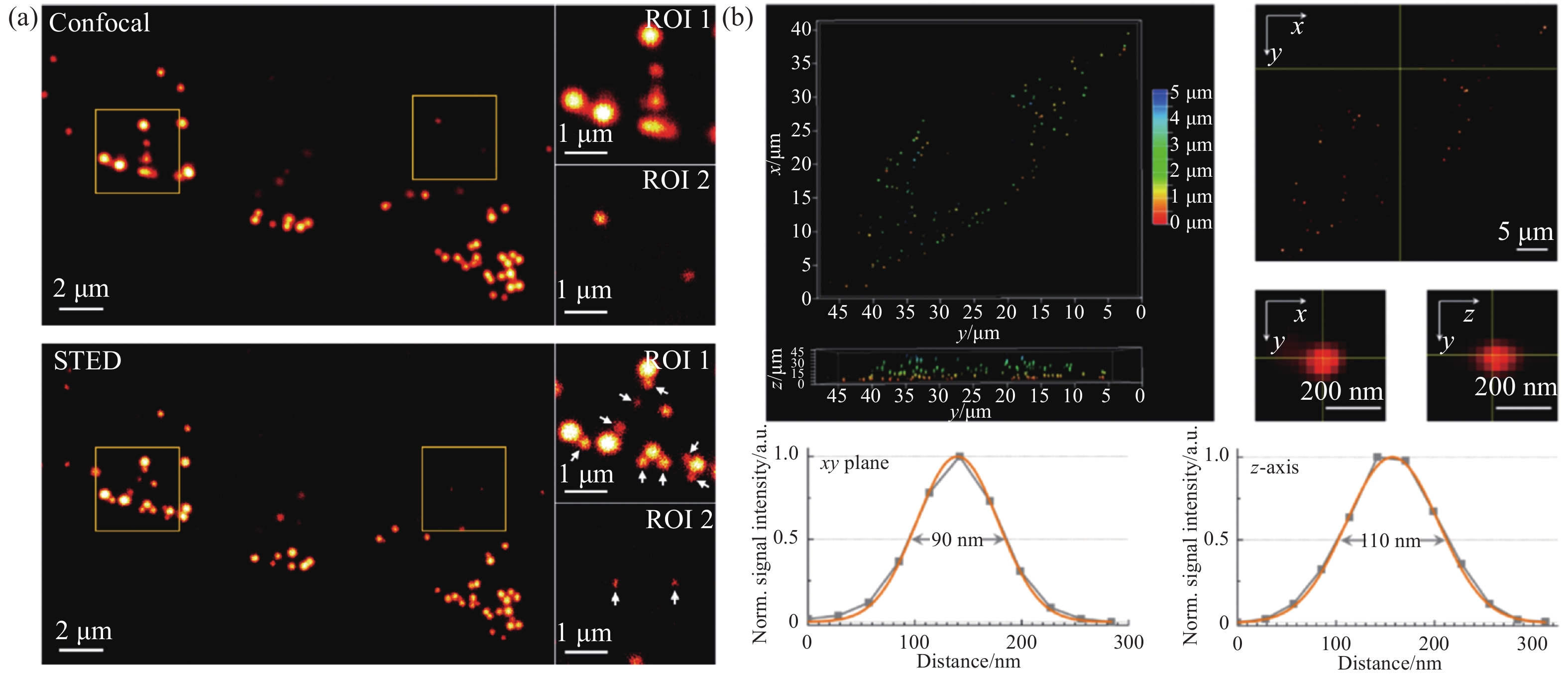
LIU G N, PENG G SH, DAI J N, et al. STED nanoscopy imaging of cellular lipid droplets employing a superior organic fluorescent probe[J]. Analytical Chemistry, 2021, 93(44): 14784-14791. doi: 10.1021/acs.analchem.1c03474
|
| [23] |
LIU G N, DAI J N, ZHOU R, et al. A distyrylbenzene-based fluorescent probe with high photostability and large Stokes shift for STED nanoscopy imaging of cellular lipid droplets[J]. Sensors and Actuators B:Chemical, 2022, 353: 131000. doi: 10.1016/j.snb.2021.131000
|
| [24] |
XU H K, ZHANG H H, LIU G, et al. Coumarin-based fluorescent probes for super-resolution and dynamic tracking of lipid droplets[J]. Analytical Chemistry, 2019, 91(1): 977-982. doi: 10.1021/acs.analchem.8b04079
|
| [25] |
O’CONNOR D, BYRNE A, BERSELLI G B, et al. Mega-stokes pyrene ceramide conjugates for STED imaging of lipid droplets in live cells[J]. Analyst, 2019, 144(5): 1608-1621. doi: 10.1039/C8AN02260G
|
| [26] |
LIU X L, XIN L, TONG Z, et al. Revealing lipid droplets evolution at nanoscale under proteohormone stimulation by a BODIPY-hexylcarbazole derivative[J]. Biosensors and Bioelectronics, 2021, 175: 112871. doi: 10.1016/j.bios.2020.112871
|
| [27] |
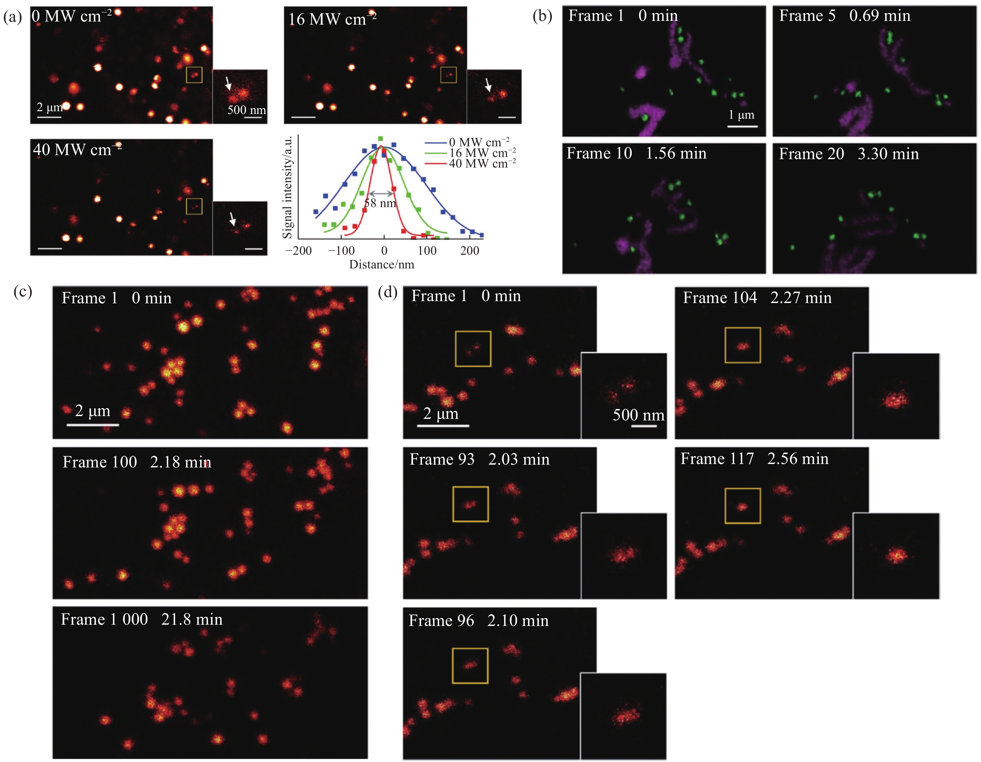
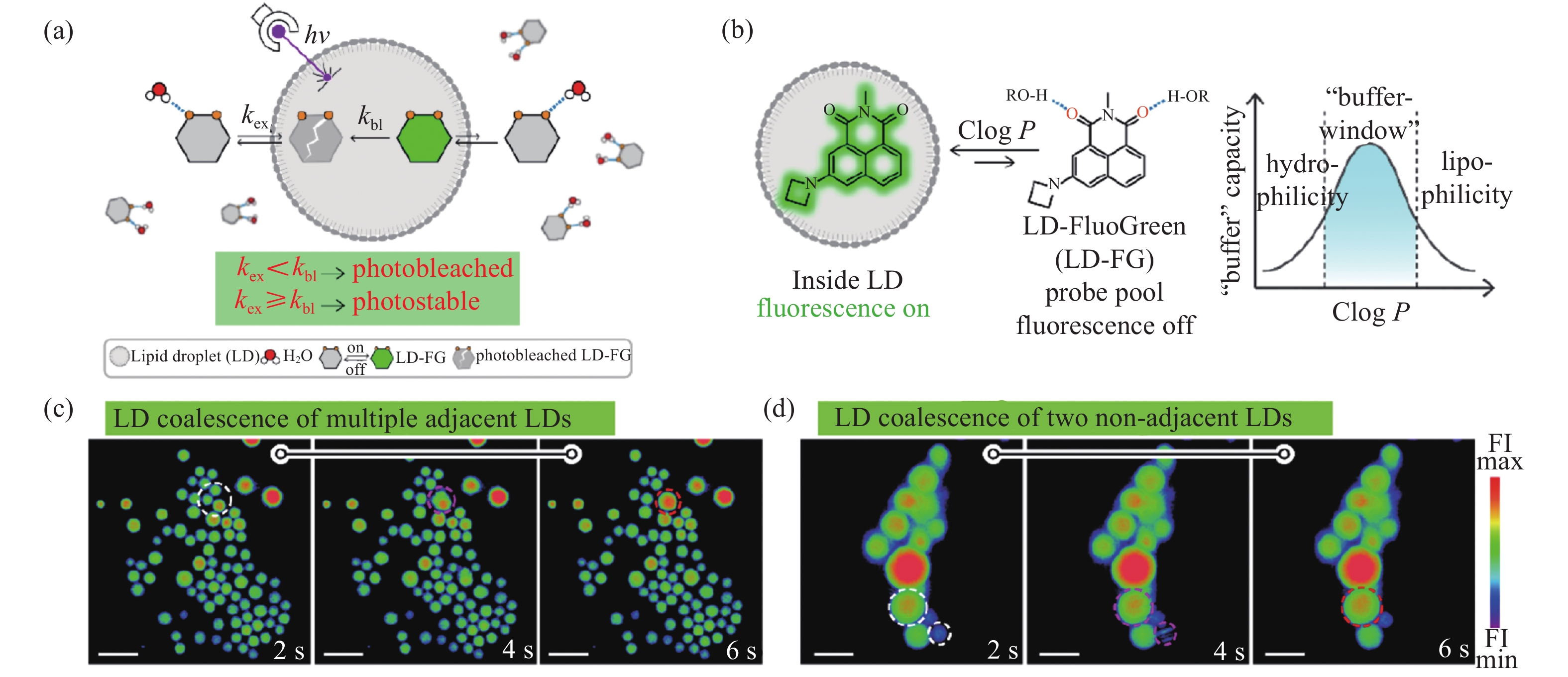
CHEN J, WANG CH, LIU W J, et al. Stable super-resolution imaging of lipid droplet dynamics through a buffer strategy with a hydrogen-bond sensitive fluorogenic probe[J]. Angewandte Chemie International Edition, 2021, 60(47): 25104-25113. doi: 10.1002/anie.202111052
|
| [28] |
WU M Y, LEUNG J K, KAM C, et al. A near-infrared AIE probe for super-resolution imaging and nuclear lipid droplet dynamic study[J]. Materials Chemistry Frontiers, 2021, 5(7): 3043-3049. doi: 10.1039/D0QM00914H
|
| [29] |
ZHENG X J, ZHU W CH, NI F, et al. A specific bioprobe for super-resolution fluorescence imaging of lipid droplets[J]. Sensors and Actuators B:Chemical, 2018, 255: 3148-3154. doi: 10.1016/j.snb.2017.09.139
|
| [30] |
ZHENG X J, ZHU W CH, NI F, et al. Simultaneous dual-colour tracking lipid droplets and lysosomes dynamics using a fluorescent probe[J]. Chemical Science, 2019, 10(8): 2342-2348. doi: 10.1039/C8SC04462G
|
| [31] |
TANG J, ROBICHAUX M A, WU K L, et al. Single-atom fluorescence switch: a general approach toward visible-light-activated dyes for biological imaging[J]. Journal of the American Chemical Society, 2019, 141(37): 14699-14706. doi: 10.1021/jacs.9b06237
|
| [32] |
WANG L SH, WANG SH CH, TANG J, et al. Oxime as a general photocage for the design of visible light photo-activatable fluorophores[J]. Chemical Science, 2021, 12(47): 15572-15580. doi: 10.1039/D1SC05351E
|
| [33] |
ADHIKARI S, BANERJEE C, MOSCATELLI J, et al. Conventional BODIPY conjugates for live-cell super-resolution microscopy and single-molecule tracking[J]. Journal of Visualized Experiments, 2020(160): 60950-60958.
|
| [34] |
YE SH, YAN W, ZHAO M J, et al. Low-saturation-intensity, high-photostability, and high-resolution STED nanoscopy assisted by CsPbBr3 quantum dots[J]. Advanced Materials, 2018, 30(23): 1800167. doi: 10.1002/adma.201800167
|
| [35] |
WANG L W, CHEN Y, PENG X, et al. Ultralow power demand in fluorescence nanoscopy with digitally enhanced stimulated emission depletion[J]. Nanophotonics, 2020, 9(4): 831-839. doi: 10.1515/nanoph-2019-0475
|
| [36] |
LI D Y, QIN W, XU B, et al. AIE nanoparticles with high stimulated emission depletion efficiency and photobleaching resistance for long-term super-resolution bioimaging[J]. Advanced Materials, 2017, 29(43): 1703643. doi: 10.1002/adma.201703643
|
| [37] |
LI D Y, NI X, ZHANG X Y, et al. Aggregation-induced emission luminogen-assisted stimulated emission depletion nanoscopy for super-resolution mitochondrial visualization in live cells[J]. Nano Research, 2018, 11(11): 6023-6033. doi: 10.1007/s12274-018-2118-5
|
| [38] |
LIU Y J, LU Y Q, YANG X S, et al. Amplified stimulated emission in upconversion nanoparticles for super-resolution nanoscopy[J]. Nature, 2017, 543(7644): 229-233. doi: 10.1038/nature21366
|
| [39] |
HUANG X SH, FAN J CH, LI L J, et al. Fast, long-term, super-resolution imaging with Hessian structured illumination microscopy[J]. Nature Biotechnology, 2018, 36(5): 451-459. doi: 10.1038/nbt.4115
|
| [40] |
ZHENG X L, DUAN R Y, LI L J, et al. Live-cell superresolution pathology reveals different molecular mechanisms of Pelizaeus-Merzbacher disease[J]. Science Bulletin, 2020, 65(24): 2061-2064. doi: 10.1016/j.scib.2020.08.016
|
| [41] |
ZHANGHAO H, CHEN X Y, LI M H, et al. Super-resolution imaging of fluorescent dipoles via polarized structured illumination microscopy[J]. Nature Communications, 2019, 10(1): 4694. doi: 10.1038/s41467-019-12681-w
|
| [42] |
GUO Y T, LI D, ZHANG S W, et al. Visualizing intracellular organelle and cytoskeletal interactions at nanoscale resolution on millisecond timescales[J]. Cell, 2018, 175(5): 1430-1442.e17. doi: 10.1016/j.cell.2018.09.057
|
| [43] |
LI D, SHAO L, CHEN B CH, et al. Extended-resolution structured illumination imaging of endocytic and cytoskeletal dynamics[J]. Science, 2015, 349(6251): aab3500. doi: 10.1126/science.aab3500
|
| [44] |
ZHAO T Y, HAO H W, WANG ZH J, et al. Multi-color structured illumination microscopy for live cell imaging based on the enhanced image recombination transform algorithm[J]. Biomedical Optics Express, 2021, 12(6): 3474-3484. doi: 10.1364/BOE.423171
|
| [45] |
WANG ZH J, ZHAO T Y, HAO H W, et al. High-speed image reconstruction for optically sectioned, super-resolution structured illumination microscopy[J]. Advanced Photonics, 2022, 4(2): 026003.
|
| [46] |
LIU ZH H, LIU J, WANG X D, et al. Fluorescent bioconjugates for super-resolution optical nanoscopy[J]. Bioconjugate Chemistry, 2020, 31(8): 1857-1872. doi: 10.1021/acs.bioconjchem.0c00320
|
| [47] |
GUI D, CHEN Y J, KUANG W B, et al. Accelerating multi-emitter localization in super-resolution localization microscopy with FPGA-GPU cooperative computation[J]. Optics Express, 2021, 29(22): 35247-35260. doi: 10.1364/OE.439976
|
| [48] |
WANG Y J, KUANG W B, SHANG M T, et al. Two-color super-resolution localization microscopy via joint encoding of emitter location and color[J]. Optics Express, 2021, 29(21): 34797-34809. doi: 10.1364/OE.440706
|
| [49] |
DU Y, WANG CH Z, ZHANG CH, et al. Computational framework for generating large panoramic super-resolution images from localization microscopy[J]. Biomedical Optics Express, 2021, 12(8): 4759-4778. doi: 10.1364/BOE.433489
|
| [50] |
HELL S W, WICHMANN J. Breaking the diffraction resolution limit by stimulated emission: stimulated-emission-depletion fluorescence microscopy[J]. Optics Letters, 1994, 19(11): 780-782. doi: 10.1364/OL.19.000780
|
| [51] |
KLAR T A, HELL S W. Subdiffraction resolution in far-field fluorescence microscopy[J]. Optics Letters, 1999, 24(14): 954-956. doi: 10.1364/OL.24.000954
|
| [52] |
BUTKEVICH A N, YU G, SIDENSTEIN S C, et al. Fluorescent rhodamines and fluorogenic carbopyronines for super-resolution STED microscopy in living cells[J]. Angewandte Chemie International Edition, 2016, 55(10): 3290-3294. doi: 10.1002/anie.201511018
|
| [53] |
BORDENAVE M D, BALZAROTTI F, STEFANI F D, et al. STED nanoscopy with wavelengths at the emission maximum[J]. Journal of Physics D:Applied Physics, 2016, 49(36): 365102. doi: 10.1088/0022-3727/49/36/365102
|
| [54] |
GÖTTFERT F, PLEINER T, HEINE J, et al. Strong signal increase in STED fluorescence microscopy by imaging regions of subdiffraction extent[J]. Proceedings of the National Academy of Sciences of the United States of America, 2017, 114(9): 2125-2130. doi: 10.1073/pnas.1621495114
|
| [55] |
SHANK N I, PHAM H H, WAGGONER A S, et al. Twisted cyanines: a non-planar fluorogenic dye with superior photostability and its use in a protein-based fluoromodule[J]. Journal of the American Chemical Society, 2013, 135(1): 242-251. doi: 10.1021/ja308629w
|
| [56] |
SHANK N I, ZANOTTI K J, LANNI F, et al. Enhanced photostability of genetically encodable fluoromodules based on fluorogenic cyanine dyes and a promiscuous protein partner[J]. Journal of the American Chemical Society, 2009, 131(36): 12960-12969. doi: 10.1021/ja9016864
|
| [57] |
OYAMA Y, MAMADA M, SHUKLA A, et al. Design strategy for robust organic semiconductor laser dyes[J]. ACS Materials Letters, 2020, 2(2): 161-167. doi: 10.1021/acsmaterialslett.9b00536
|
| [58] |
MICHIE M S, GÖTZ R, FRANKE C, et al. Cyanine conformational restraint in the far-red range[J]. Journal of the American Chemical Society, 2017, 139(36): 12406-12409. doi: 10.1021/jacs.7b07272
|
| [59] |
ZHOU R, CUI Y Y, DAI J N, et al. A red-emissive fluorescent probe with a compact single-benzene-based skeleton for cell imaging of lipid droplets[J]. Advanced Optical Materials, 2020, 8(13): 1902123. doi: 10.1002/adom.201902123
|
| [60] |
YANG X S, YANG ZH G, WU ZH Y, et al. Mitochondrial dynamics quantitatively revealed by STED nanoscopy with an enhanced squaraine variant probe[J]. Nature Communications, 2020, 11(1): 3699. doi: 10.1038/s41467-020-17546-1
|
| [61] |
LIU Y J, DING Y CH, ALONAS E, et al. Achieving λ/10 resolution CW STED nanoscopy with a Ti: sapphire oscillator[J]. PLoS One, 2012, 7(6): e40003. doi: 10.1371/journal.pone.0040003
|
| [62] |
BIANCHINI P, HARKE B, GALIANI S, et al. Single-wavelength two-photon excitation-stimulated emission depletion (SW2PE-STED) superresolution imaging[J]. Proceedings of the National Academy of Sciences of the United States of America, 2012, 109(17): 6390-6393. doi: 10.1073/pnas.1119129109
|
| [63] |
GUSTAFSSON M G L. Surpassing the lateral resolution limit by a factor of two using structured illumination microscopy: short communication[J]. Journal of Microscopy, 2000, 198(2): 82-87. doi: 10.1046/j.1365-2818.2000.00710.x
|
| [64] |
GUSTAFSSON M G L. Nonlinear structured-illumination microscopy: wide-field fluorescence imaging with theoretically unlimited resolution[J]. Proceedings of the National Academy of Sciences of the United States of America, 2005, 102(37): 13081-13086. doi: 10.1073/pnas.0406877102
|
| [65] |
骆清铭, 张镇西. 生物医学光子学[M]. 北京: 人民卫生出版社, 2018.
LUO Q M, ZHANG Z X. Biomedical Photonics[M]. Beijing: People's Medical Publishing House, 2018. (in Chinese)
|
| [66] |
刘志贺, 吴长锋. 超分辨率成像荧光探针材料应用进展[J]. 中国光学,2018,11(3):344-362. doi: 10.3788/co.20181103.0344
LIU ZH H, WU CH F. Advances in application of materials of super-resolution imaging fluorescent probe[J]. Chinese Optics, 2018, 11(3): 344-362. (in Chinese) doi: 10.3788/co.20181103.0344
|
| [67] |
SPAHN C, GRIMM J B, LAVIS L D, et al. Whole-cell, 3D, and multicolor STED imaging with exchangeable fluorophores[J]. Nano Letters, 2019, 19(1): 500-505. doi: 10.1021/acs.nanolett.8b04385
|
| [68] |
BETZIG E, PATTERSON G H, SOUGRAT R, et al. Imaging intracellular fluorescent proteins at nanometer resolution[J]. Science, 2006, 313(5793): 1642-1645. doi: 10.1126/science.1127344
|
| [69] |
BRIEKE C, ROHRBACH F, GOTTSCHALK A, et al. Light-controlled tools[J]. Angewandte Chemie International Edition, 2012, 51(34): 8446-8476. doi: 10.1002/anie.201202134
|
| [70] |
LI W H, ZHENG G H. Photoactivatable fluorophores and techniques for biological imaging applications[J]. Photochemical &Photobiological Sciences, 2012, 11(3): 460-471.
|
| [71] |
SENGUPTA P, VAN ENGELENBURG S B, LIPPINCOTT-SCHWARTZ J. Superresolution imaging of biological systems using photoactivated localization microscopy[J]. Chemical Reviews, 2014, 114(6): 3189-3202. doi: 10.1021/cr400614m
|
| [72] |
NANI R R, GORKA A P, NAGAYA T, et al. Near-IR light-mediated cleavage of antibody-drug conjugates using cyanine photocages[J]. Angewandte Chemie International Edition, 2015, 54(46): 13635-13638. doi: 10.1002/anie.201507391
|
| [73] |
GWOSCH K C, PAPE J K, BALZAROTTI F, et al. MINFLUX nanoscopy delivers 3D multicolor nanometer resolution in cells[J]. Nature Methods, 2020, 17(2): 217-224. doi: 10.1038/s41592-019-0688-0
|

Figures(12) / Tables(1)


 DownLoad: