


-
摘要: 金属纳米颗粒的等离激元共振引起的局域场增强效应,对显微成像、光谱学、半导体器件、非线性光学等诸多领域都具有极大的应用潜力。尤其是在光学纳米材料领域,通过亚波长金属纳米颗粒与电介质的组合引起局域场增强效应,提高了纳米材料的光学性能,并促进纳米材料在光学领域的应用。本文主要综述几种常见纳米结构所产生的局域场增强效应及其应用,详细介绍并总结了金属纳米材料的不同结构参数与局域场增强的关系及局域场增强在非线性光学、光谱学、半导体器件等领域的应用。未来,随着对金属纳米材料的研究愈发深入,局域场增强的应用将更加广泛,这将对诸多领域的发展产生重要影响。Abstract: Local field enhancement(LFE) based on the plasmon resonance characteristics of metal nanoparticles has great potential in many fields such as microscopy, spectroscopy, semiconductor devices and nonlinear optics. Especially in the field of optical nanomaterials, local field enhancement effect can be produced by the combination of sub-wavelength metal nanoparticles and dielectrics to improve the optical properties of nanomaterials and promote the application of nanomaterials in the field of optics. In this paper, the local field enhancement effect of several common nanostructures and their applications is mainly reviewed. The relationship between different structural parameters of metal nanomaterials and the local field enhancement and the application of local field enhancement in nonlinear optics, spectroscopy, semiconductor devices are introduced and summarized. It is foreseeable that in the future, as the research on metal nanomaterials progresses, the application of localized field enhancement will be more extensive, which have a significant impact on the development of many fields.
-
Key words:
- surface plasmon resonance /
- local field enhancement /
- nanostructures /
- nonlinear effect
-

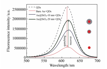
图 1 不同厚度SiO2外壳包裹的小金纳米颗粒对CdTe量子点荧光强度的影响
Figure 1. Effect of small gold nanoparticles with different thickness of SiO2 coating on the fluorescence spectra of CdTe QDs

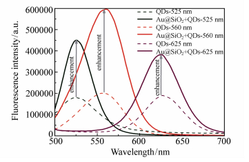
图 2 SiO2包裹的大金纳米颗粒对不同发射波长的CdTe量子点的荧光增强
Figure 2. Effect of silicon-coated large gold nanoparticles on the fluorescence enhancement of CdTe QDs with different emission wavelength

图 3 包裹不同SiO2壳厚度(a)0 nm, (b)3 nm, (c)14 nm, (d)38 nm的AuNRs@SiO2的TEM图像
Figure 3. TEM images for Au NRs coated with (a)0 nm, (b)3 nm, (c)14 nm, and (d)38 nm silica shells

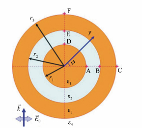
图 4 “核壳”金纳米结构的几何构型图
Figure 4. Geometrical configuration of the core-shell gold nanostructures

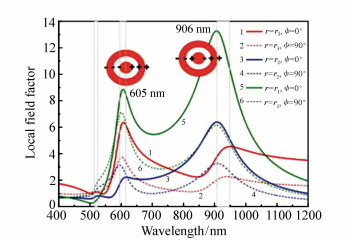
图 5 不同位置的局域场因子光谱图
Figure 5. Spectra of different positions local field factor r1=10 nm, r2=15 nm, r3=20 nm, ε2=4.2, ε4=1.8

图 6 局域电场因子与插入金球的波长和半径的函数关系:(a)C点, (b)F点, (c)B点, (d)E点, (e)A点, (f)D点
Figure 6. Local electric field factor in the gold-dielectric-gold nanoshells as a function of wavelength and radius of the inserted gold sphere:(a)at point C, (b)at point F, (c)at point B, (d)at point E, (e)at point A, and (f)at point D

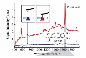
图 8 Ag涂覆的尖端探针测量BCB产生的拉曼光谱
Figure 8. Tip-enhanced Raman spectra of brilliant cresyl blue BCB dispersed on a glass support measured with a silver-coated AFM probe

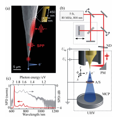
图 9 纳米聚焦表面等离子体激元(SPP)的实验示意图
Figure 9. Experimental schemes of nanofocused surface plasmon Excitations(SPP). (a)SEM image of a gold tip with a grating coupler 20 μm away from the apex with illustration of SPP nanofocusing triggering ultrafast electron emission. (b)Corresponding electron pulse imaging setup using an ultrashort 5 fs laser system for plasmon excitation. (c)Normalized spectral power density(SPD) of the ultra-broadband spectrum of the laser system

图 10 飞秒激光作用尖端金涂层产生的四波混频信号成像(a)Au-Si近场FWM图像,图中“S1,S2,S3”是对应的“热点”,(b)同一时刻的原子力显微镜图像,(c)双脉冲激发,对应于τ=0 fs 8.2 fs, 16.4 fs不同脉冲间延迟下,同一位置的四波混频图像,(d)对“S1”及“S2”处四波混频强度随去相位时间变化的模拟图,(e)沿a图(蓝色)和b图(黑色)中的白色虚线提取的FWM(蓝色)信号及AFM(黑色)形貌图(彩图见电子版)
Figure 10. Femtosecond FWM nanoimaging of coherent plasmon dynamics in gold. (a)Near-field FWM image of a Si-Au step, showing 'hotspots' S1, S2 and S3. (b)Simultaneously acquired AFM topography. (c)FWM images of the same region with two-pulse excitation, corresponding to an inter-pulse delay of τ=0 fs(top), 8.2 fs(middle) and 16.4 fs(bottom), demonstrating evolution of the relative intensities in spots S1, S2 and S3. (d)FWM intensity in S1 and S2 for the three delays, showing variation in dephasing time T2, with simulation for T2=16 fs(black solid line) and T2=10 fs(red solid line). (e)Line profiles of FWM signal(blue), showing ~50 nm spatial resolution, and AFM topography(black), extracted from (a) and (b) along the white dashed lines(color figures see electronic version)

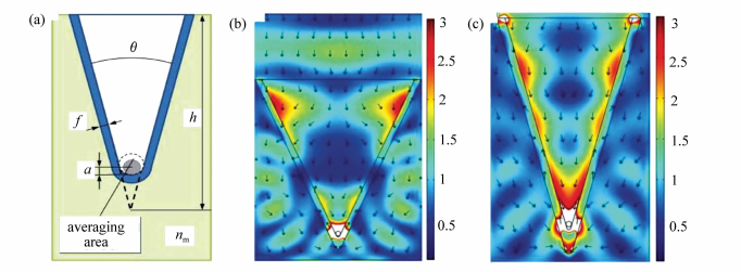
图 11 (a) 紫外区局域场增强的“V”形纳米结构几何形状。颜色表示电场分布,箭头表示能流方向; (b)nm=2.1,f=17 nm,θ=50°,h=480 nm,a=9 nm;(c)nm=1.7,f=20 nm,θ=32°,h=560 nm,a=9 nm,“V”形槽内介质的介电常数为1
Figure 11. (a)The geometry of V-shaped nanostructure for local field enhancement in UV region. Distribution of the electric field in the resonator(color) and direction of the power flow(arrows) for two sets of parameters: (b)nm=2.1, f=17 nm, θ=50°, h=480 nm, a=9 nm; (c)nm=1.7, f=20 nm, θ=32°, h=560 nm, and a=9 nm. Dielectric constant of the medium inside the V-groove equals to 1. (For interpretation of the references to color in this figure legend, the reader is referred to the web version of this article)

图 12 (a) f=17 nm,a=9 nm时局域场强度|E(h, θ)|随深度h和孔径角θ的变化关系。(b)θ=50°,h=480 nm时局域场强度|E(f, a)|随金属膜厚度f和尖端圆角半径a的变化关系。电解质的折射率nm=2.1,“V”形槽内介质折射率为1
Figure 12. (a)Dependence of the local field enhancement |E(h, θ)| on depth h and aperture angle θ, for f=17 nm and a=9 nm. (b)Dependence of the local field enhancement |E(f, a)| on the thickness of the metal film f and the fillet radius a, for θ=50°, h=480 nm. Refractive index of the dielectric medium is equal nm=2.1, refractive index of the medium incide the V-groove is 1

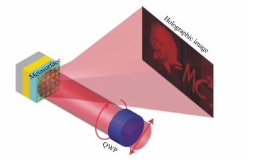
图 13 在圆偏振入射光束下的基于反射纳米棒的CGH的图示,圆偏振入射光束通过四分之一波片(QWP)落在表面上,反射光束在远场中形成全息图像
Figure 13. Illustration of the reflective nanorod-based CGH under a circularly polarized incident beam. The circularly polarized incident beam, which is converted from a linearly polarized one by passing through a quarter wave plate(QWP), falls on the metasurface. The reflected beam forms the holographic image in the far field

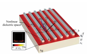
图 14 基于与银膜耦合的银纳米片的等离子体超表面的示意图
Figure 14. Schematic illustration of the plasmonic metasurface based on silver nanostripes coupled to a silver film

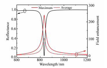
图 15 线性超表面与入射波长的反射率(黑线)和场增强(红线)分布。红色实线和虚线分别描绘了局域场增强和空间平均场增强的相对值
Figure 15. Reflectance (black line) and field enhancement (red lines) distributions of the linear metasurface versus the incident wavelength. The red solid and dotted lines depict the local maximum and the spatially averaged field enhancement, respectively

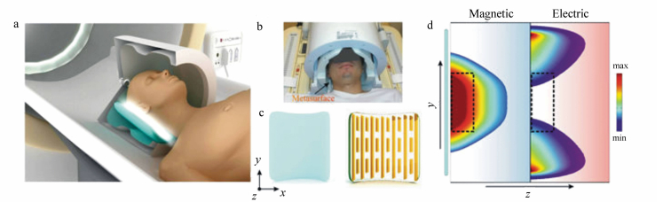
图 16 超材料的几何结构与近场处磁场和电场分布模拟图,(a)磁共振成像装置切割示意图,(b)发射(外部)和多元件接收线圈阵列(内部)的体内实验的照片,(c)高介电常数电介质基片(左)与其金属结构(右)组成的超表面结构,(d)数值计算出的磁(左)和电(右)场在超表面附近的映射(显示为蓝色矩形)
Figure 16. Structural geometry of the metamaterial and simulation diagram of near field magnetic and electric field distributions. (a)Schematic of the MRI setup with a cut-out for better visualization of the setup. (b)A photograph of the in-vivo experiment including the transmit(outer) and multi-element receive coil array(inner). (c)Artist's view of the hybrid metasurface, including high permittivity dielectric substrate(left) combined with its metallic structure(right). (d)Numerically calculated magnetic(left) and electric(right) field maps in vacuum near the metasurfaces(shown as a blue rectangle)

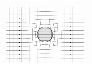
图 18 在球状掺杂物附近主体区域局部电场的集中
Figure 18. Field lines and equipotential surfaces inside and outside a spherical inclusion particle, plotted for εi > εh

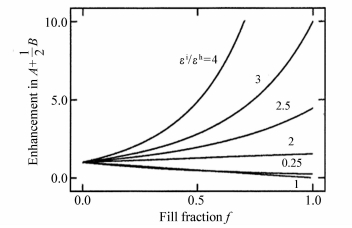
图 19 介质的非线性系数A+(1/2)B预测χ(3)增强与线性掺杂粒子填充分数的函数关系
Figure 19. Predicted enhancement in the nonlinear coefficient A+(1/2)B, which is proportional to χ(3), as a function of the volume fill fraction of nonlinear material for linear inclusion particles embedded in a nonlinear host material

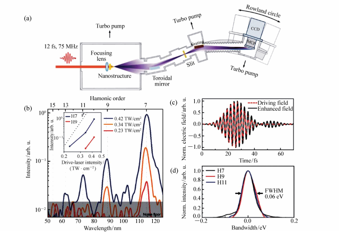
图 20 金属-蓝宝石纳米结构产生高次谐波示意图,(a)用于产生极紫外和光谱测量的实验装置,(b)通过测量极紫外光谱得到高次谐波(HHG)峰,(c)FDTD模拟入射激光场与蓝宝石尖端增强场的时间曲线,(d)HHG峰的归一化曲线,带宽表示每个峰值的光子能量除以其谐波阶次。FWHM:半峰宽
Figure 20. High-harmonic generation from the metal-sapphire nanostructure. (a)Overall hardware configuration for extreme ultraviolet generation and spectrum measurement. (b)Measured extreme ultraviolet spectra showing HHG peaks. (c)FDTD-simulated temporal profile of the enhanced field at the sapphire tip for the incident laser field. (d)Normalized profiles of measured HHG peaks. The bandwidth represents the photon energy spread of each peak divided by its harmonic order. FWHM: full-width at half-maximum
表 1 有无AuNRs@SiO2的器件光伏参数
Table 1. Photovoltaic parameters for devices with and without AuNRs@SiO2
SiO2 thickness/nm Voc/V Jsc/(mA·cm-2) FF PCE/% Rs/(Ω·cm2) Rsh/(Ω·cm2) ref. 0.74 16.5 0.60 7.52 10.1 801 3±0.6 0.74 21.2 0.60 9.55 7.4 923 14±2 0.74 19.1 0.60 8.53 8.7 322 38±5 0.74 18.5 0.60 8.25 7.8 435  下载: 导出CSV
-
[1] ZIELINSKI M, WINTER S, KOLKOWSKI R, et al.. Nanoengineering the second order susceptibility in semiconductor quantum dot heterostructures[J]. Opt. Express, 2011, 19(7):6657-6670. doi: 10.1364/OE.19.006657 [2] WANG SH W, QIAN J, HE S L, et al.. Three-photon luminescence of gold nanorods and its applications for high contrast tissue and deep in vivo brain imaging[J]. Ivyspring. Theranostics, 2015, 5(3):251-266. doi: 10.7150/thno.10396 [3] ZHUANG Z Y, YANG Q, ZHANG Z M, et al.. A highly selective fluorescent probe for hydrogen peroxide and its applications in living cells[J]. Journal of Photochemistry and Photobiology A:Chemistry, 2017, 344:8-14. doi: 10.1016/j.jphotochem.2017.04.009 [4] MANDAL K, JANA D, GHORAI B, et al.. Fluorescent imaging probe from nanoparticle made of aie molecule[J]. Phys. Chem. C, 2016, 120(9):5196-5206. doi: 10.1021/acs.jpcc.5b12682 [5] XU Q, HEO CH, JIN A K, et al.. A selective imidazoline-2-thione-bearing two-photon fluorescent probe for hypochlorous acid in mitochondria[J]. Anal. Chem., 2016, 88(12):6615-6620. doi: 10.1021/acs.analchem.6b01738 [6] KAURANEN M, ZAYATS A V. Nonlinear plasmonics[J]. Nature Photonics, 2012, 6(11):737-748. doi: 10.1038/nphoton.2012.244 [7] JASSIM N M, WANG K, HAN X, et al.. Plasmon assisted enhanced second-harmonic generation in single hybrid Au/ZnS nanowires[J]. Optical Materials, 2017, 64:257-261. doi: 10.1016/j.optmat.2016.11.034 [8] 王马华, 朱光平, 居勇峰, 等.纳米氧化锌中三光子吸收与倍频效应致光辐射特性[J].发光学报, 2015, 36(6):617-622. http://www.opticsjournal.net/Abstract.htm?id=OJ150625000093iOlRnUWANG M H, ZHU G P, JU Y F, et al.. Emission characteristics of crown-like ZnO nanocrystals induced by three-photon absorption and second harmonic generation effect[J]. Chinese J. Luminescence, 2015, 36(6):617-622.(in Chinese) http://www.opticsjournal.net/Abstract.htm?id=OJ150625000093iOlRnU [9] 朱华, 颜振东, 詹鹏, 等.局域表面等离激元诱导的三次谐波增强效应[J].物理学报, 2013, 62(17): 178104. doi: 10.7498/aps.62.178104ZHU H, YAN ZH D, ZHAN P, et al.. Third harmonic generation enhancement effect induced by local surface plasmon[J]. Acta Phys. Sin., 2013, 62(17):178104.(in Chinese) doi: 10.7498/aps.62.178104 [10] W YE, W ZHANG, S WANG, et al.. Effect of sapphire substrate on the localized surface plasmon resonance of aluminum triangular nanoparticles[J]. Optics Communications, 2017, 395:175-182. doi: 10.1016/j.optcom.2016.01.089 [11] KUMAR A, DIXIT T, PALANI I A, et al.. Utilization of surface plasmon resonance of Au/Pt nanoparticles for highly photosensitive ZnO nanorods network based plasmon field effect transistor[J]. Physica E:Low-dimensional Systems and Nanostructures, 2017, 93:97-104. doi: 10.1016/j.physe.2017.06.005 [12] AGHLARA H, ROSTAMI R, MAGHOUL A, et al.. Noble metal nanoparticle surface plasmon resonance in absorbing medium[J]. Optik-International Journal for Light and Electron Optics, 2015, 126(4):417-420. doi: 10.1016/j.ijleo.2013.12.089 [13] SAFONOV A L, SULYAEVA V S, TIMOSHENKO N I, et al.. Deposition of thin composite films consisting of fluoropolymer and silver nanoparticles having surface plasmon resonance[J]. Thin Solid Films, 2016, 603:313-316. doi: 10.1016/j.tsf.2016.02.030 [14] YAN L, YAN Y, XU L, et al. Large range localized surface plasmon resonance of Ag nanoparticles films dependent of surface morphology[J]. Applied Surface Science, 2016, 367:563-568. doi: 10.1016/j.apsusc.2016.01.238 [15] 薛彬, 孔祥贵, 王丹, 等.785 nm激光诱导银纳米三角片聚集表面增强拉曼散射效应[J].中国光学, 2014, 7(1):118-123. http://www.chineseoptics.net.cn/CN/abstract/abstract9104.shtmlXUE B, KONG X G, WANG D, et al.. SERS effect of aggregation of silver nanoprisms induced by 785 nm laser[J]. Chinese Optics, 2014, 7(1):118-123.(in Chinese) http://www.chineseoptics.net.cn/CN/abstract/abstract9104.shtml [16] 封昭, 周骏, 陈栋, 等.基于金/银纳米三明治结构SERS特性的超灵敏前列腺特异性抗原检测[J].发光学报, 2015, 36(9):1064-1070. http://www.opticsjournal.net/Abstract.htm?id=OJ151022000066C0FbIeFENG ZH, ZHOU J, CHEN D, et al.. Hypersensitization immunoassay of prostate-specific antigen based on SERS of sandwich-type Au/Ag nanostructure[J]. Chinese J. Luminescence, 2015, 36(9):1064-1070.(in Chinese) http://www.opticsjournal.net/Abstract.htm?id=OJ151022000066C0FbIe [17] 李晓坤, 张友林, 孔祥贵.Ag纳米粒子聚集体的SiO2包覆及其SERS效应[J].发光学报, 2014, 35(7):853-857. http://www.opticsjournal.net/abstract.htm?id=OJ140218000123B9EbHdLI X K, ZHANG Y L, KONG X G. Aggregation of Ag nanoparticles coated with silica and its SERS effect[J]. Chinese J. Luminescence, 2014, 35(7):853-857.(in Chinese) http://www.opticsjournal.net/abstract.htm?id=OJ140218000123B9EbHd [18] SÖNNICHSEN C, ALIVISATOS A. Gold nanorods as novel nonbleaching plasmon-based orientation sensors for polarized single-particle microscopy[J]. Nano Lett., 2005, 5(2):301-304. doi: 10.1021/nl048089k [19] MURPHY C J, SAU T K, GOLE A M, et al.. Anisotropic metal nanoparticles:synthesis, assembly, and optical applications[J]. Phys. Chem. B, 2005, 109(29):13857-13870. doi: 10.1021/jp0516846 [20] JIA K, YUAN L, ZHOU X, et al.. One-pot synthesis of Au/Ag bimetallic nanoparticles to modulate the emission of CdSe/CdS quantum dots[J]. RSC Adv., 2015, 5:58163-58170. doi: 10.1039/C5RA08933F [21] ZHU J, CHANG H, LI J J, et al.. Using silicon-coated gold nanoparticles to enhance the fluorescence of CdTe quantum dot and improve the sensing ability of mercury(Ⅱ)[J]. Molecular and Biomolecular Spectroscopy, 2017. http://www.ncbi.nlm.nih.gov/pubmed/28709143 [22] ZHANG R, ZHOU Y, PENG L, et al.. Influence of SiO2 shell thickness on power conversion efficiency in plasmonic polymer solar cells with Au nanorod@SiO2core-shell structures[J]. Scientific Reports, 2016, 6:25036. doi: 10.1038/srep25036 [23] ZHU J, REN Y, ZHAO S, et al.. The effect of inserted gold nanosphere on the local field enhancement of gold nanoshell[J]. Materials Chemistry and Physics, 2012, 133(2-3):1060-1065. doi: 10.1016/j.matchemphys.2012.02.016 [24] JIANG N, DMITRY KUROUSKI, POZZI E A, et al.. Tip-enhanced Raman spectroscopy:from concepts to practical applications[J]. Chemical Physics Letters, 2016, 659:16-24. doi: 10.1016/j.cplett.2016.06.035 [25] GAURAV SHARMA, VOLKER DECKERT, et al.. Tip-enhanced Raman scattering-Targeting structure-specific surface characterization for biomedical samples[J]. Advanced Drug Delivery Reviews, 2015, 89:42-56. doi: 10.1016/j.addr.2015.06.007 [26] JUNG Y, CHEN H, TONG L, et al.. Imaging gold nanorods by plasmon-resonance-enhanced four wave mixing[J]. Journal of Physical Chemistry C, 2009, 113(7):2657-2663. doi: 10.1021/jp810852c [27] MVLLER M, KRAVTSOV V, PAARMANN A, et al.. A nanofocused plasmon-driven sub-10 femtosecond electron point source[J]. ACS Photonics, 2016, 3(4):611-619. doi: 10.1021/acsphotonics.5b00710 [28] KRAVTSOV V, ULBRICHT R, ATKIN J M, et al.. Plasmonic nanofocused four-wave mixing for femtosecond near-field imaging[J]. Nature Nanotechnology, 2016, 11(5):459-464. doi: 10.1038/nnano.2015.336 [29] SHALIN A S, SUKHOV S V, KRASNUK A E, et al.. Plasmonic nanostructures for local field enhancement in the UV region[J]. Photonics and Nanostructures-Fundamentals and Applications, 2014, 12(1):2-8. https://www.sciencedirect.com/science/article/pii/S1569441013000709 [30] ZHENG G, M HLENBERND H, KENEY M, et al.. Metasurface holograms reaching 80% efficiency[J]. Nature Nanotechnology, 2015, 10(4):308-312. doi: 10.1038/nnano.2015.2 [31] JIN B, ARGYROPOULOS C. Enhanced four-wave mixing with nonlinear plasmonic metasurfaces[J]. Scientific Reports, 2016, 6:28746. doi: 10.1038/srep28746 [32] SCHMIDT R, SLOBOZHANYUK A, BELOV P, et al.. Flexible and compact hybrid metasurfaces for enhanced ultra high field in vivo magnetic resonance imaging[J]. Scientific Reports, 2017, 7:1678. doi: 10.1038/s41598-017-01932-9 [33] JE SIPE, RW BOYD, Nanocomposite materials for nonlinear optics based on local field effects[J]. Springer Berlin Heidelberg, 2002, 82(4):1-19. http://www.springerlink.com/content/jrm27m1h4magmky0 [34] RW BOYD, JE SIPE, et al.. Nonlinear optical properties of nanocomposite materials[J]. Pure & Applied Optics Journal of the European Optical Society Part A, 1996, 5(5):505. https://www.researchgate.net/profile/Robert_Boyd4/publication/231132905_Nonlinear_optical_properties_of_nanocomposite_materials/links/542d5df00cf29bbc126d2b16.pdf?inViewer=true&disableCoverPage=true&origin=publication_detail [35] GHIMIRE S, DICHIARA A D, SISTRUNK E, et al.. Observation of high-order harmonic generation in a bulk crystal[J]. Nature Physics, 2011, 7(2):138-141. doi: 10.1038/nphys1847 [36] HAN S, KIM H, YONG W K, et al.. High-harmonic generation by field enhanced femtosecond pulses in metal-sapphire nanostructure[J]. Nature Communications, 2016, 7:13105. doi: 10.1038/ncomms13105 [37] VAMPA G, GHAMSARI B G, HAMMOND T J, et al.. Plasmon-enhanced high-harmonic generation from silicon[J]. Nature Physics, 2017, 13:659-662. doi: 10.1038/nphys4087 [38] 帕拉斯·N·普拉萨德.纳米光子学[M].西安:西安交通大学出版社, 2010.PARAS N. PRASAD. Nanophotonics[M]. Xi'an:Xi'an Jiaotong University Press, 2010. [39] ZHU W, ESTEBAN R, BORISOV A G, et al.. Quantum mechanical effects in plasmonic structures with subnanometre gaps[J]. Nature Communications, 2016, 7:11495. doi: 10.1038/ncomms11495 -

 下载:
下载:


计量
- 文章访问数: 4290
- HTML全文浏览量: 1073
- PDF下载量: 819
- 被引次数: 0
