


-
摘要: 本文设计了一种用于婴幼儿视网膜筛查的广域眼底相机。文中对该系统所包含的照明系统、成像系统的设计方法进行了探讨。首先,根据James Polans宽视场人眼模型和婴儿眼解剖学数据,建立了婴儿眼模型。接着,提出新的锥形光纤方案用于大视场照明。最后,重点介绍了广域眼底相机的成像系统(包括接触镜、中继物镜)的设计方法。设计实例表明:广域眼底相机的视场可以达到130°,对眼底的物方分辨率可以达到10 μm。设计结果符合眼底成像设备国家标准YY0634-2008,满足婴幼儿视网膜筛查的要求。Abstract: A wide-area fundus camera used for screening the retinae of infants was designed. In this paper, the design methods of the device’s illuminating and imaging systems were investigated. Based on James Polans’ wide-field human eye model and a set of ophthalmic anatomy data, an infant eye model was established. Then, a tapered fiber scheme was proposed for wide area illumination. Finally, the design method of a wide-area fundus camera imaging system, including the contact lens and relay lens, is introduced. The design example shows that the Field Of View (FOV) of the wide-area fundus camera can reach 130°, and the object resolution of the fundus can reach 10 μm. The design results meet the national standards YY0634-2008 for fundus imaging equipment and meet the requirements for infant retina screening.
-
Key words:
- optical design /
- wide-area retinal imaging /
- medical optics
-

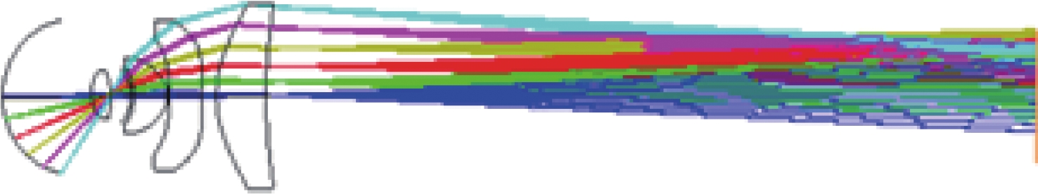
图 4 锥形光纤照明范围的仿真结果
Figure 4. Simulation results of illumination range of tappered fiber
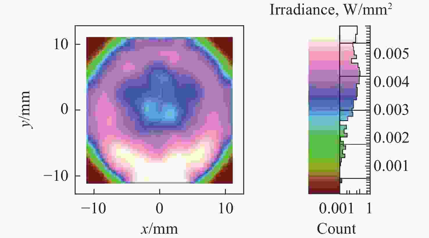
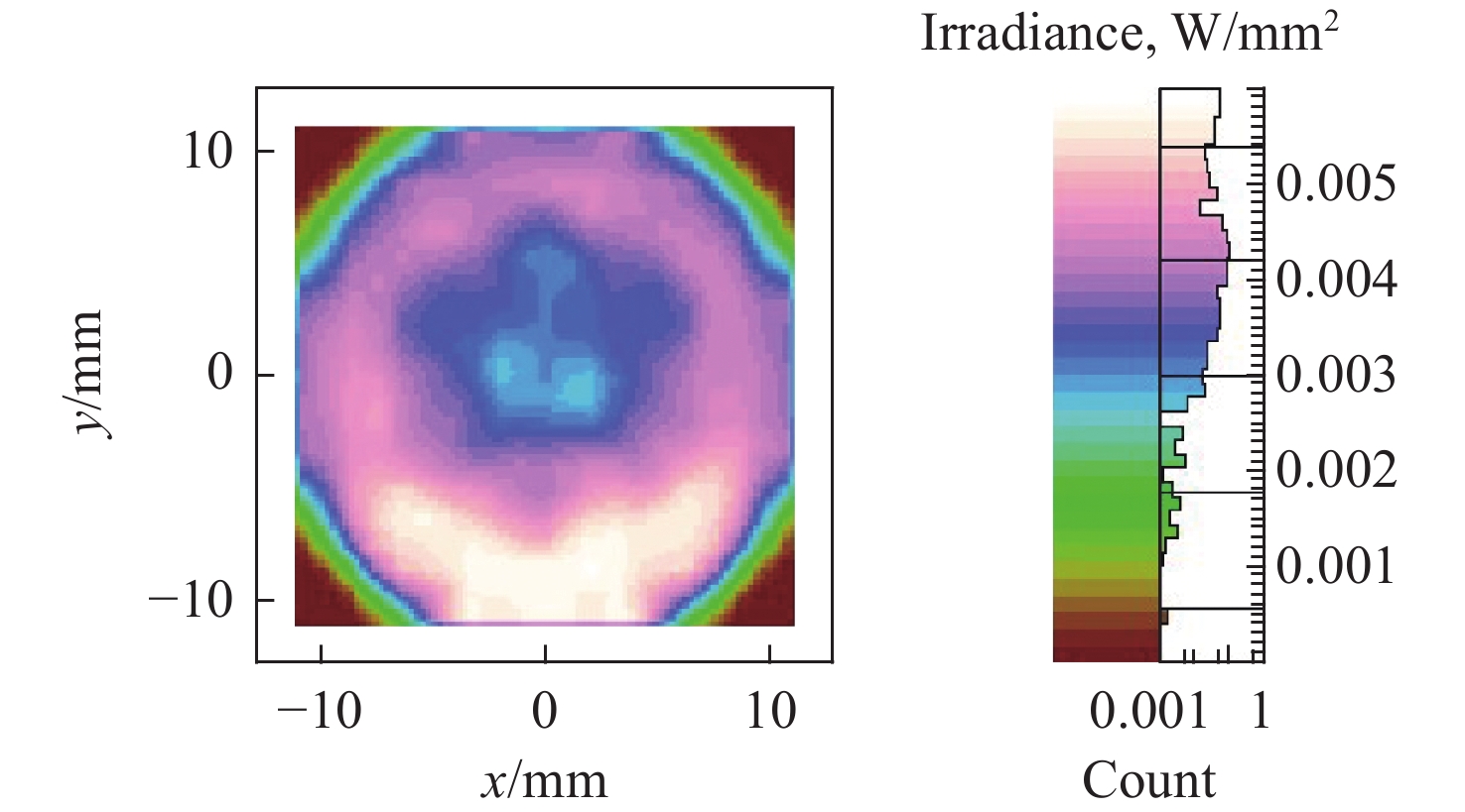
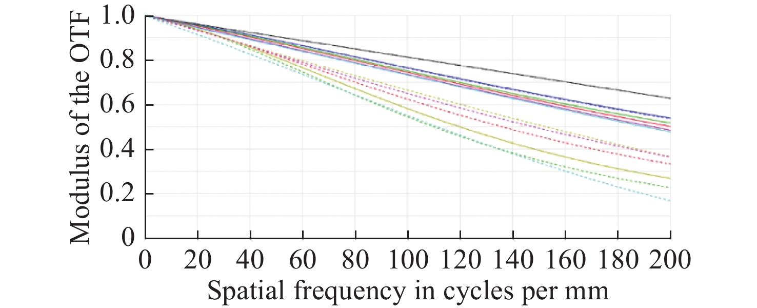
图 5 锥形光纤照度均匀性的仿真结果
Figure 5. Simulation results of illumination uniformity of tappered fiber

图 12 原图(a)和利用本文广域眼底相机光学系统(b)所得的仿真图
Figure 12. (a) Original image and (b) simulation image obtained by optical system in proposed wide-area fundus camera
表 1 婴儿眼结构参数
Table 1. Parameters of an infant eye
解剖学数据 眼模型参数 曲率半径/mm 厚度/mm 材料 曲率半径/mm 厚度/mm 材料 角膜前表面 < 7.62 < 0.4 Nd ≈ 1.38 4.428 0.32 参考James Polans模型 角膜后表面 < 6.2 − − 3.519 − − 房水 − < 2.9 Nd ≈ 1.34 − 2.10 参考James Polans模型 晶状体前表面 未知 2.9 ~ 4 Nd ≈ 1.38~1.44 6.633 2.90 参考James Polans模型 晶状体后表面 未知 − − −3.738 − − 玻璃体 − 12 ~ 13 Nd ≈ 1.34 − 12.90 参考James Polans模型 视网膜 < 8.5 − − −7.533 − − 视轴总长 17.2 ~ 19.8 18.22  下载: 导出CSV
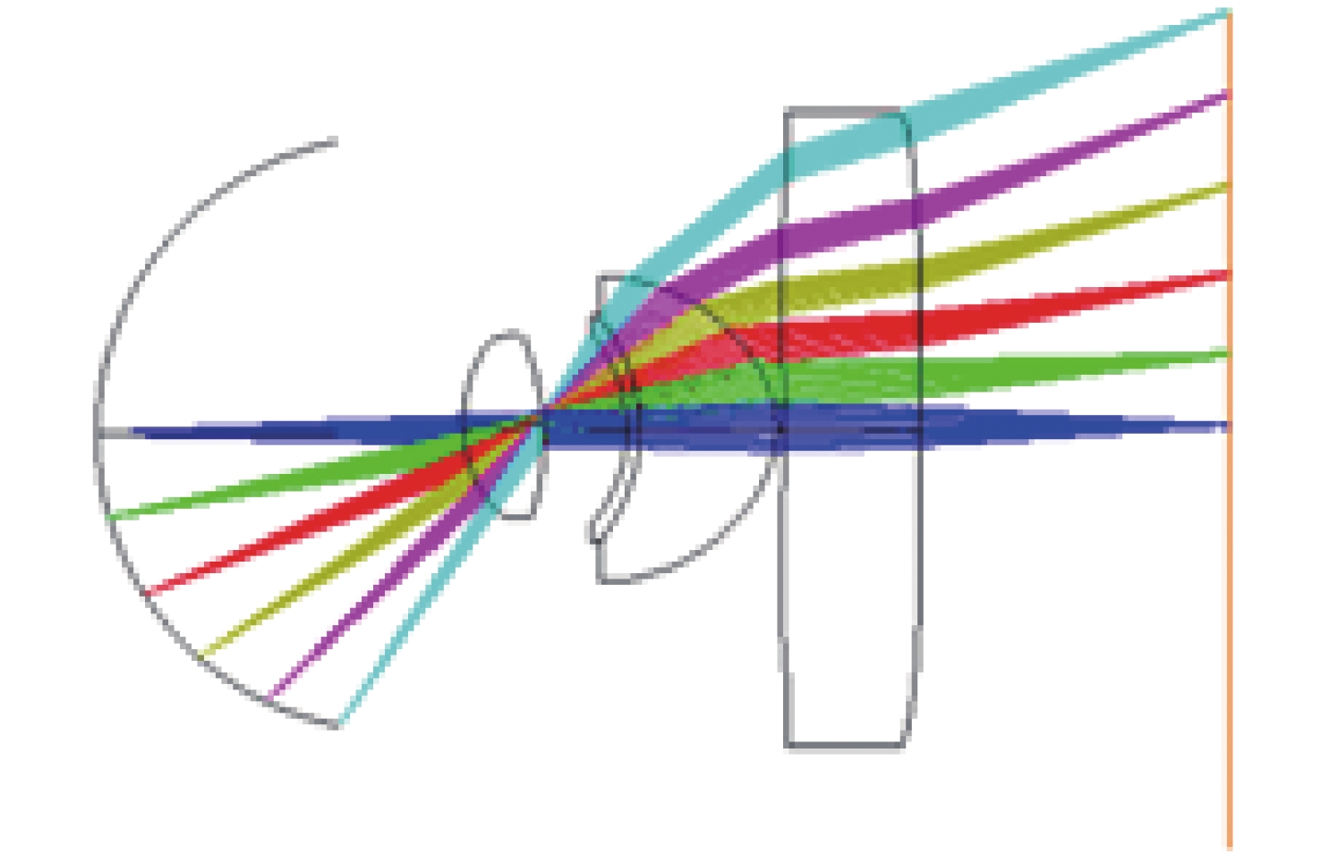
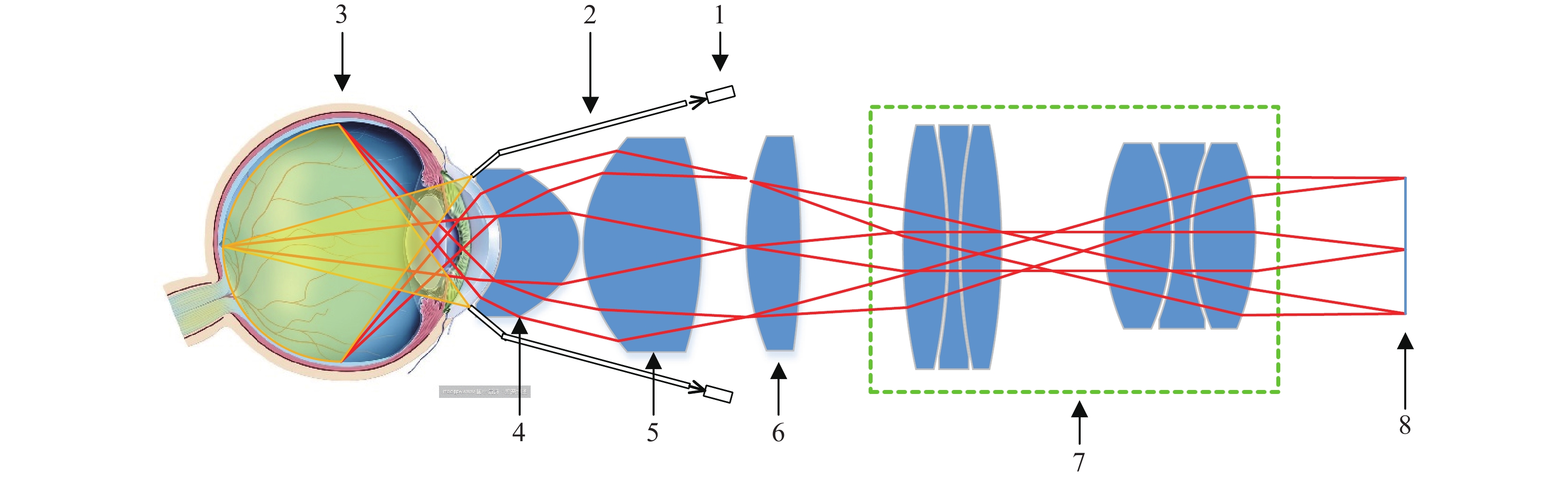
表 2 成像系统结构参数
Table 2. Parameters of imaging system
Surf Radius (mm) Thickness (mm) Glass nd vd 1 −4.428 3.456 1.67 55 2 −2.767 1.728 3 −37.936 3.456 1.85 40 4 125.526 1.441 5 9.230 5.186 1.67 55 6 49.036 23.042 7 40.087 5.733 1.76 27 8 −35.656 10.325 9 −5.948 3.853 1.73 2 10 −9.848 3.075 11 −11.061 4.542 1.85 24 12 −44.707 1.097 13 −96.658 3.469 1.57 56 14 −13.075 2.880 15 23.308 2.951 1.59 61 16 −45.371 1.395 17 9.583 4.928 1.83 37 18 6.156 2.452 19 14.406 2.386 1.59 61 20 −5.937 1.726 1.92 21 21 −18.279 0.319 22 3.961 2.304 1.59 61 23 9.911 3.517 IMG
下载: 导出CSV
-
[1] 赵堪兴, 杨培增. 眼科学[M]. 8版. 北京: 人民卫生出版社, 2013.ZHAO K X, YANG P Z. Ophthalmology[M]. 8th ed. Beijing: People's Medical Publishing House, 2013. (in Chinese) [2] 李蕊, 刘永基, 王肇圻. 基于个体眼光学结构的波前眼镜设计[J]. 中国光学,2012,5(5):512-519.LI R, LIU Y J, WANG ZH Q. Design of wavefront-guided lens based on individual eye optical model[J]. Chinese Optics, 2012, 5(5): 512-519. (in Chinese) [3] 王植.广域数字眼底成像关键技术研究[D]. 南京: 南京航空航天大学, 2017.WANG ZH. Research on key technique of wide-area digital fundus imaging technology[D]. Nanjing: Nanjing University of Aeronautics and Astronautics, 2017. (in Chinese) [4] 姚凤莹, 沈建新, 陈华. 婴幼儿视网膜广域成像关键技术研究[J]. 光学与光电技术,2018,16(6):63-70.YAO F Y, SHEN J X, CHEN H. Study on key techniques of wide-area retinal imaging for infant[J]. Optics &Optoelectronic Technology, 2018, 16(6): 63-70. (in Chinese) [5] 王业顺, 沈建新. 广域视网膜成像技术研究[J]. 应用光学,2018,39(1):35-39. doi: 10.5768/JAO201839.0101007WANG Y SH, SHEN J X. Research of wide-area retinal imaging technique[J]. Journal of Applied Optics, 2018, 39(1): 35-39. (in Chinese) doi: 10.5768/JAO201839.0101007 [6] 沈冬玲, 张光华, 马娟, 等. 应用RetCam3行足月新生儿视网膜筛查的临床研究[J]. 中国实用医药,2019,14(22):26-27.SHEN D L, ZHANG G H, MA J, et al. Clinical study of RetCam3 for full-term neonatal retinal screening[J]. China Practical Medicine, 2019, 14(22): 26-27. (in Chinese) [7] 张国明, 曾键, 黄丽娜, 等. 广角数码儿童视网膜成像系统引导下激光光凝治疗早产儿视网膜病变[J]. 中华眼底病杂志,2008,24(1):17-19.ZHANG G M, ZENG J, HUANG L N, et al. Wide-field digital pediatric retinal imaging system-assisted photocoagulation for retinopathy of prematurity[J]. Chinese Journal of Ocular Fundus Diseases, 2008, 24(1): 17-19. (in Chinese) [8] 李秋平, 黄俊谨, 陈颖, 等. 早产儿视网膜病变筛查分析[J]. 中华眼科杂志,2012,48(10):903-907. doi: 10.3760/cma.j.issn.0412-4081.2012.10.010LI Q P, HUANG J J, CHEN Y, et al. Retinopathy of prematurity screening in 2185 premature infants[J]. Chinese Journal of Ophthalmology, 2012, 48(10): 903-907. (in Chinese) doi: 10.3760/cma.j.issn.0412-4081.2012.10.010 [9] MANIVANNAN A, PLSKOVA J, FARROW A, et al. Ultra-wide-field fluorescein angiography of the ocular fundus[J]. American Journal of Ophthalmology, 2005, 140(3): 525-527. doi: 10.1016/j.ajo.2005.02.055 [10] PURBRICK R M J, IZADI S, GUPTA A, et al. Comparison of optomap ultrawide-field imaging versus slit-lamp biomicroscopy for assessment of diabetic retinopathy in a real-life clinic[J]. Clinical Ophthalmology, 2014, 8: 1413-1417. [11] DEHOOG E, SCHWIEGERLING J. Fundus camera systems: a comparative analysis[J]. Applied Optics, 2009, 48(2): 221-228. doi: 10.1364/AO.48.000221 [12] 李灿.新型眼底相机的设计与研制[D]. 长春: 中国科学院研究生院(长春光学精密机械与物理研究所), 2014.LI C. Design and fabrication of new type of fundus camera[D]. Changchun: Changchun Institute of Optics, Fine Mechanics and Physics, Chinese Academy of Sciences, 2014. (in Chinese) [13] 王晶, 崔恩坤. 大视场曲面复眼光学系统设计[J]. 中国光学,2014,7(6):969-974. doi: 10.3788/CO.20140706.0969WANG J, CUI E K. Design of large FOV curved artificial compound eye with freeform lens[J]. Chinese Optics, 2014, 7(6): 969-974. (in Chinese) doi: 10.3788/CO.20140706.0969 [14] POLANS J, JAEKEN B, MCNABB R P, et al. Wide-field optical model of the human eye with asymmetrically tilted and decentered lens that reproduces measured ocular aberrations[J]. Optica, 2015, 2(2): 124-134. doi: 10.1364/OPTICA.2.000124 [15] 李秋明, 郑广瑛. 眼科应用解剖学[M]. 郑州大学出版社, 2010.LI Q M, ZHENG G Y. Applied Anatomy of Ophthalmology[M]. Zhengzhou: Zhengzhou University Press, 2010. (in Chinese) [16] 中华人民共和国国家质量监督检验检疫总局, 中国国家标准化管理委员会. GB/T 7247.9-2016 激光产品的安全 第9部分: 非相干光辐射最大允许照射量[S]. 北京: 中国标准出版社, 2017.AQSIQ, Standardization Administration of China. GB/T 7247.9-2016 Safety of laser products—Part 9: Compilation of maximum permissible exposure to incoherent optical radiation[S]. Beijing: Standards Press of China, 2017. (in Chinese) [17] 何远清, 刘永基, 翟奕. 成像角膜曲率计的光学设计[J]. 中国光学,2014,7(6):956-961. doi: 10.3788/CO.20140706.0956HE Y Q, LIU Y J, ZHAI Y. Optical design of imaging keratometer[J]. Chinese Optics, 2014, 7(6): 956-961. (in Chinese) doi: 10.3788/CO.20140706.0956 [18] 程少园.视网膜血管的液晶自适应光学成像系统设计[D]. 长春: 中国科学院研究生院(长春光学精密机械与物理研究所), 2010.CHENG SH Y. Design of liquid crystal adaptive optical system for fundus blood vessel imaging[D]. Changchun: Changchun Institute of Optics, Fine Mechanics and Physics, Chinese Academy of Sciences, 2010. (in Chinese) [19] 国家食品药品监督管理局. YY 0634-2008 眼科仪器 眼底照相机[S]. 北京: 中国标准出版社, 2009.State Food and Drug Administration. YY 0634-2008 Ophthalmic instruments—fundus cameras[S]. Beijing: Standards Press of China, 2009. (in Chinese) -

 下载:
下载:


计量
- 文章访问数: 4410
- HTML全文浏览量: 1602
- PDF下载量: 430
- 被引次数: 0
