Enhancing the fluorescence emission by flexible metal-dielectric-metal structures
doi: 10.37188/CO.2021-0084
-
摘要: 增强荧光发射可以提高荧光检测灵敏度、提高LED的亮度,在提高发光器件性能方面具有重要意义。由于金属结构在增强局域场、增强荧光发射方面具有很好效果,而柔性电介质材料具有灵活的可弯曲性特性,本文提出一种由金属-电介质-金属(MDM)组成的柔性结构以增强荧光发射。利用时域有限差分方法系统研究了该结构对量子点定向发射增强的影响。理论计算表明柔性MDM结构局部起伏和弧度对荧光增强起促进作用,且可以使位于结构中心位置量子点的量子效率增强约7倍。此外,还可以改变电介质的折射率和厚度从而实现目标波长的可调谐性。实验结果表明该柔性MDM结构对荧光增强有一定的促进作用,这一发现对未来的显示技术和柔性发光器件都有很大的价值,对高效柔性器件的开发应用具有一定的指导意义。Abstract: The technology of enhancing fluorescence emission can increase the sensitivity of fluorescence detection and the brightness of Light Emitting Diodes (LEDs), and is of great significance in improving the performance of light-emitting devices. Since the metal structure has a good effect in enhancing the local field and fluorescence emission, and the flexible dielectric material has flexible bendability characteristics, on the basis of above, we propose a flexible structure composed of Metal-Dielectric-Metal (MDM) to enhance the fluorescence emission. The influence of the structure on the directional emission enhancement of quantum dots is systematically studied by using the finite difference time domain method. Theoretical calculations show that the local undulations and arcs of the flexible MDM structure can promote fluorescence enhancement and increase the quantum efficiency of the quantum dots located at the center of the structure by about 7 times. They can alao change the refractive index and thickness of the dielectric to achieve the tunability of the target wavelength. At the same time, the experimental results shows that the flexible MDM structure does have a positive effect on the fluorescence enhancement. This discovery is valuable for future display technologies and flexible light-emitting devices. It is of certain guiding significance for the development and application of high-efficiency flexible devices.
-
Key words:
- fluorescence enhancement /
- flexible structure /
- directional emission /
- tunable wavelength
-
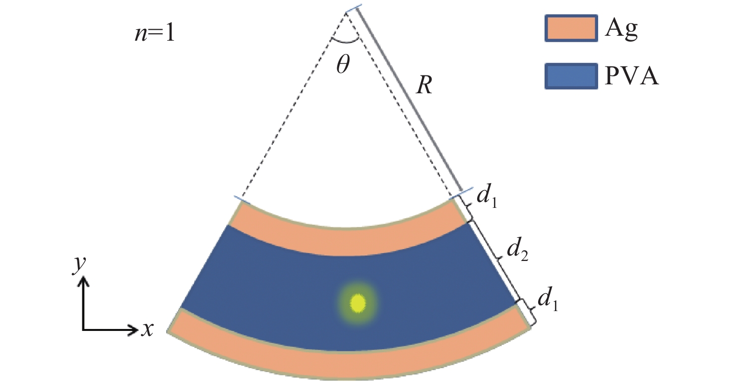
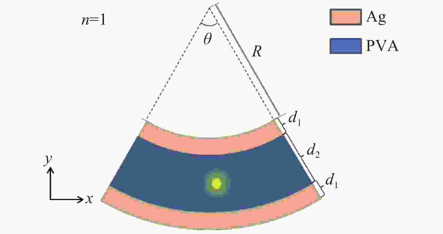
图 1 银和PVA组成的MDM结构模型示意图,其中橙色区域代表银膜,其厚度为d1;蓝色区域代表电介质PVA,其厚度为d2,上层银膜内半径为R,结构所对应圆心角为θ
Figure 1. Schematic diagram of the MDM structure model composed of silver and PVA, in which: the orange area represents the silver film with a thickness of d1; the blue area represents the dielectric PVA with a thickness of d2, the inner radius of the upper silver film is R, and the central angle corresponding to the structure is θ
-
[1] WANG Z B, HELANDER M G, QIU J, et al. Unlocking the full potential of organic light-emitting diodes on flexible plastic[J]. Nature Photonics, 2011, 5(12): 753-757. doi: 10.1038/nphoton.2011.259 [2] KIM W, KWON S, LEE S M, et al. Soft fabric-based flexible organic light-emitting diodes[J]. Organic Electronics, 2013, 14(11): 3007-3013. doi: 10.1016/j.orgel.2013.09.001 [3] HUANG W B, ZHANG X J, YANG T CH, et al. A mechanically bendable and conformally attachable polymer membrane microlaser array enabled by digital interference lithography[J]. Nanoscale, 2020, 12(12): 6736-6743. doi: 10.1039/C9NR10970F [4] CHOUDHURY S D, BADUGU R, RAY K, et al. Steering fluorescence emission with metal-dielectric-metal structures of Au, Ag, and Al[J]. The Journal of Physical Chemistry C, 2013, 117(30): 15798-15807. doi: 10.1021/jp4051066 [5] GRANADOS J A O, THANGARASU P, SINGH N, et al. Tetracycline and its quantum dots for recognition of Al3+ and application in milk developing cells bio-imaging [J]. Food Chemistry, 2019, 278: 523-532. doi: 10.1016/j.foodchem.2018.11.086 [6] CHEN W L, LONG K D, YU H, et al. Enhanced live cell imaging via photonic crystal enhanced fluorescence microscopy [J]. Analyst, 2014, 139(22): 5954-5963. doi: 10.1039/C4AN01508H [7] MCHUGH K J, JING L H, BEHRENS A M, et al. Biocompatible semiconductor quantum dots as cancer imaging agents[J]. Advanced Materials, 2018, 30(18): 1706356. doi: 10.1002/adma.201706356 [8] BHASIKUTTAN A C, MOHANTY J, NAU W M, et al. Efficient fluorescence enhancement and cooperative binding of an organic dye in a supra-biomolecular host-protein assembly[J]. Angewandte Chemie International Edition, 2007, 46(22): 4120-4122. doi: 10.1002/anie.200604757 [9] NANDIMATH M, BHAJANTRI R F, NAIK J. Spectroscopic and color chromaticity analysis of rhodamine 6G dye-doped PVA polymer composites for color tuning applications[J]. Polymer Bulletin, 2021, 78(8): 4569-4592. doi: 10.1007/s00289-020-03332-y [10] NGO Q M, HO Y L D, PUGH J R, et al. Enhanced UV/blue fluorescent sensing using metal-dielectric-metal aperture nanoantenna arrays[J]. Current Applied Physics, 2018, 18(7): 793-798. doi: 10.1016/j.cap.2018.04.007 [11] LI D Y, ZHOU D L, XU W, et al. Plasmonic photonic crystals induced two-order fluorescence enhancement of blue perovskite nanocrystals and its application for high-performance flexible ultraviolet photodetectors[J]. Advanced Functional Materials, 2018, 28(41): 1804429. doi: 10.1002/adfm.201804429 [12] YAN Y ZH, ZENG Y, WU Y, et al. Ten-fold enhancement of ZnO thin film ultraviolet-luminescence by dielectric microsphere arrays[J]. Optics Express, 2014, 22(19): 23552-23564. doi: 10.1364/OE.22.023552 [13] JIANG J J, XIE Y B, LIU ZH Y, et al. Amplified spontaneous emission via the coupling between Fabry-Perot cavity and surface plasmon polariton modes[J]. Optics Letters, 2014, 39(8): 2378-2381. doi: 10.1364/OL.39.002378 [14] REN Y, LU Y H, ZANG T Y, et al. Fluorescence emission mediated by metal-dielectric-metal fishnet metasurface: spatially selective excitation and double enhancement[J]. Chinese Journal of Chemical Physics, 2019, 32(3): 349-356. doi: 10.1063/1674-0068/cjcp1807182 [15] CHOUDHURY S D, BADUGU R, NOWACZYK K, et al. Tuning fluorescence direction with plasmonic metal–dielectric–metal substrates[J]. The Journal of Physical Chemistry Letters, 2013, 4(1): 227-232. doi: 10.1021/jz301867b [16] JUNG B Y, KIM N Y, LEE C H, et al. Optical properties of Fabry-Perot microcavity with organic light emitting materials[J]. Current Applied Physics, 2001, 1(2-3): 175-181. doi: 10.1016/S1567-1739(01)00006-2 [17] UDDIN S Z, TANVIR M R, TALUKDER M A. A proposal and a theoretical analysis of an enhanced surface plasmon coupled emission structure for single molecule detection[J]. Journal of Applied Physics, 2016, 119(20): 204701. doi: 10.1063/1.4952576 [18] CHOUDHURY S D, BADUGU R, RAY K, et al. Directional emission from metal-dielectric-metal structures: effect of mixed metal layers, dye location, and dielectric thickness[J]. The Journal of Physical Chemistry C, 2015, 119(6): 3302-3311. doi: 10.1021/jp512174w [19] PALIK E D. Handbook of Optical Constants of Solids[M]. Orlando: Academic Press, 1985.. [20] LU G W, ZHANG T Y, LI W Q, et al. Single-molecule spontaneous emission in the vicinity of an individual gold nanorod[J]. The Journal of Physical Chemistry C, 2011, 115(32): 15822-15828. doi: 10.1021/jp203317d [21] CHOU R Y, LU G W, SHEN H M, et al. A hybrid nanoantenna for highly enhanced directional spontaneous emission[J]. Journal of Applied Physics, 2014, 115(24): 244310. doi: 10.1063/1.4885422 [22] GRYCZYNSKI I, MALICKA J, NOWACZYK K, et al. Effects of sample thickness on the optical properties of surface plasmon-coupled emission[J]. The Journal of Physical Chemistry B, 2004, 108(32): 12073-12083. doi: 10.1021/jp0312619 -


 下载:
下载:
