Luminescence enhancement mechanism of Er3+ ions by Ag@SiO2 core-shell nanostructure in tellurite glass
-
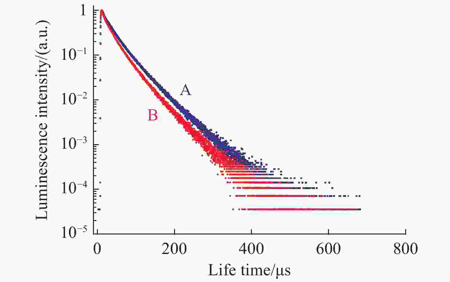
摘要: 本研究首次把预先制备好的Ag@SiO2纳米核壳结构成功地引进到碲化物发光玻璃70TeO2-25ZnO-5La2O3-0.5Er2O3体内,发现(A) Ag(1.6×10⁻⁶ mol /L)@SiO2(40 nm) @Er3+(0.5%):铒碲发光玻璃相对于样品(B) Er3+(0.5%):铒碲发光玻璃的可见光与红外光的激发光谱强度的最大增强依次为149.0%与161.5%,可见光与红外光的发光光谱强度则依次最大增强了155.2%与151.6%,同时还发现样品(A)相对于样品(B)的寿命显著变长。由于Ag@SiO2的表面等离子体吸收峰恰好位于546.0 nm,它与铒离子的发光峰546.0 nm完全共振,因此,Ag@SiO2对铒碲发光玻璃的发光共振增强作用显著。由于银的纳米核壳结构与玻璃的制作具有分步实现的优点,它既能成功控制Ag@SiO2的尺寸,而且在Ag@SiO2@Er: 铒碲发光玻璃的制作过程中还具有可操作性强的优点,同时价格也更加便宜。在保证银不被氧化的前提下,还可控制稀土离子发光中心与银的表面等离子体之间的距离,因此能够成功地减少背向能量反传递。上述优点促成了Ag@SiO2纳米核壳结构表面等离子体有效加强了Ag@SiO2@Er3+:铒碲发光玻璃的常规光致发光强度。
-
关键词:
- Ag@SiO2纳米核壳结构 /
- 发光的增强作用 /
- 表面等离离子体
Abstract: In this paper, we introduce a prefabricabed Ag@SiO2 nanostructure directly into tellurite luminescence glass composed of 70TeO2-25ZnO-5La2O3-0.5Er2O3. We find that the maximum enhancement of visible and infrared excitation spectra intensity of (A) Ag (1.6×10⁻⁶ mol/L)@SiO2(40 nm) @Er3+ (0.5%): tellurite glass relative to (B) Er3+ (0.5%): tellurite glass is about 149.0% and 161.5%, respectively. Their maximum enhancement of visible and infrared luminescence spectra intensity is 155.2% and 151.6%, respectively. We also find that sample (A) has a larger lifespan compared to sample (B). Because the surface plasmon absorption peak of Ag@SiO2 is located at 546.0 nm, it completely resonates with the luminescence peak of erbium ions which are also at 546.0 nm. Therefore, the resonance enhancement action of Ag@SiO2 on the luminescence of erbium-doped tellurite luminescence glass is significant. Thanks to the advantages of the step-by-step realization of the silver nano core-shell structure and the production of glass, it can successfully and smoothly control the size of Ag@SiO2. It also has the advantage of strong operability in the manufacturing process of Ag@SiO2@Er: telluride luminescence glass. Its costs are also minor. Moreover, it can not only ensure that the silver is not oxidized, but it can also successfully control the distance between the rare earth ion luminescence center and the silver surface plasma. It can also successfully reduce the back energy transfer, which allows the silver surface plasma to more effectively enhance the intensity of photo-luminescence. -
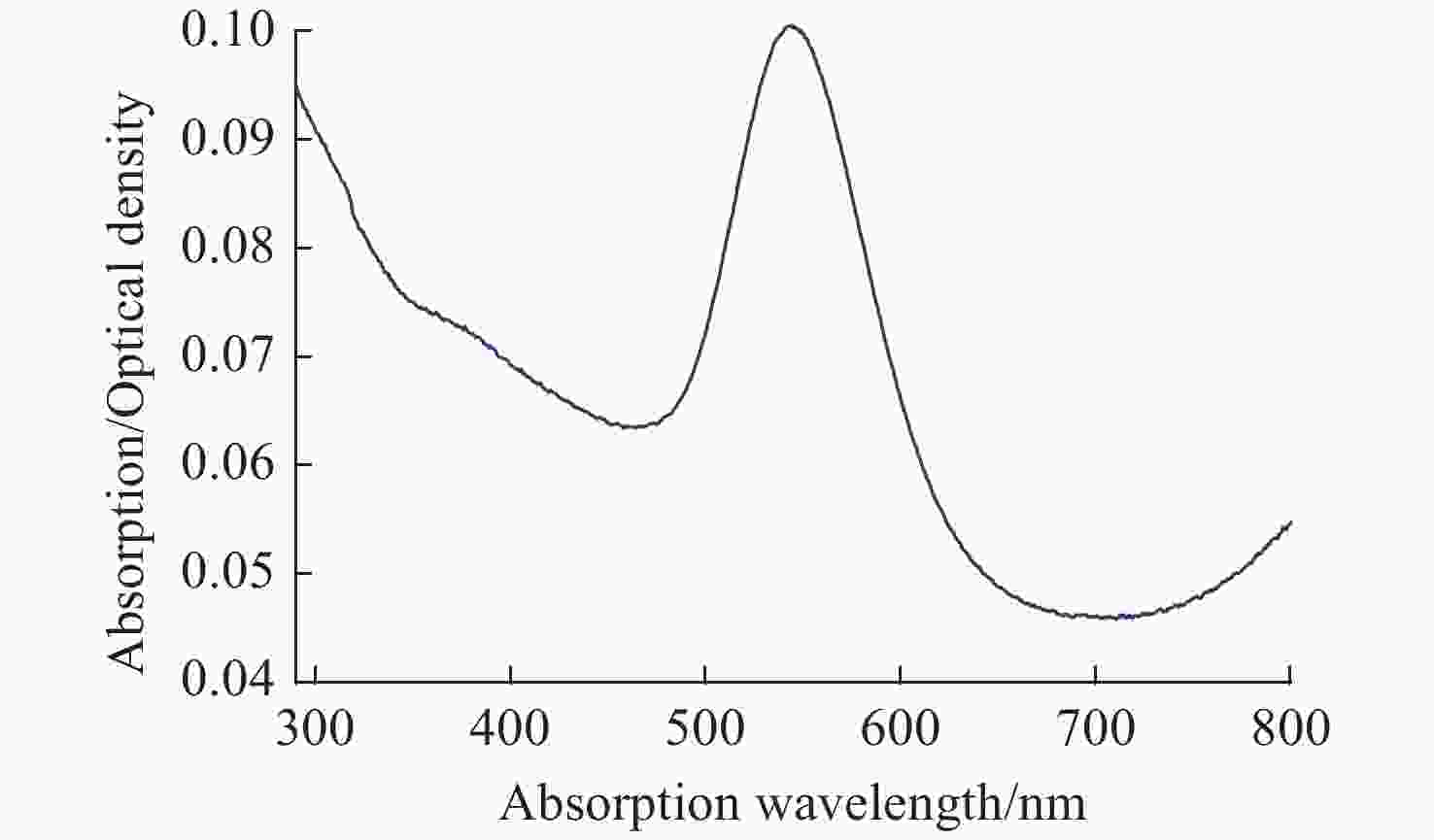
图 2 270~1800 nm波长范围内(A) Ag(1.6×10⁻⁶ mol /L) @SiO2(40 nm)@Er3+(0.5%): 铒碲发光玻璃样品(A蓝线)与(B) Er3+(0.5%): 铒碲发光玻璃样品(B红线)的吸收光谱
Figure 2. Absorption spectrum of (A) Ag(1.6×10⁻⁶ mol/L)@SiO2(40 nm) @Er3+(0.5%):TeZnLa glass (blue line A) and (B) Er(0.5%):TeZnLa glass (red line B) when measured from 270 nm to 1800 nm
图 4 Er3+Ag0: TeZnLa样品的能级结构与表面等离子体增强发光过程的示意图。蓝线、红线与绿线依次代表吸收、发光与共振散射增强过程。
Figure 4. Schematic diagram of the energy level structure and luminescence enhancement process induced by the surface plasmon of the Er3+Ag0: TeZnLa sample. The blue line, red line and green lines represent the absorption, luminescence and resonant scatter enhancement process respectively.
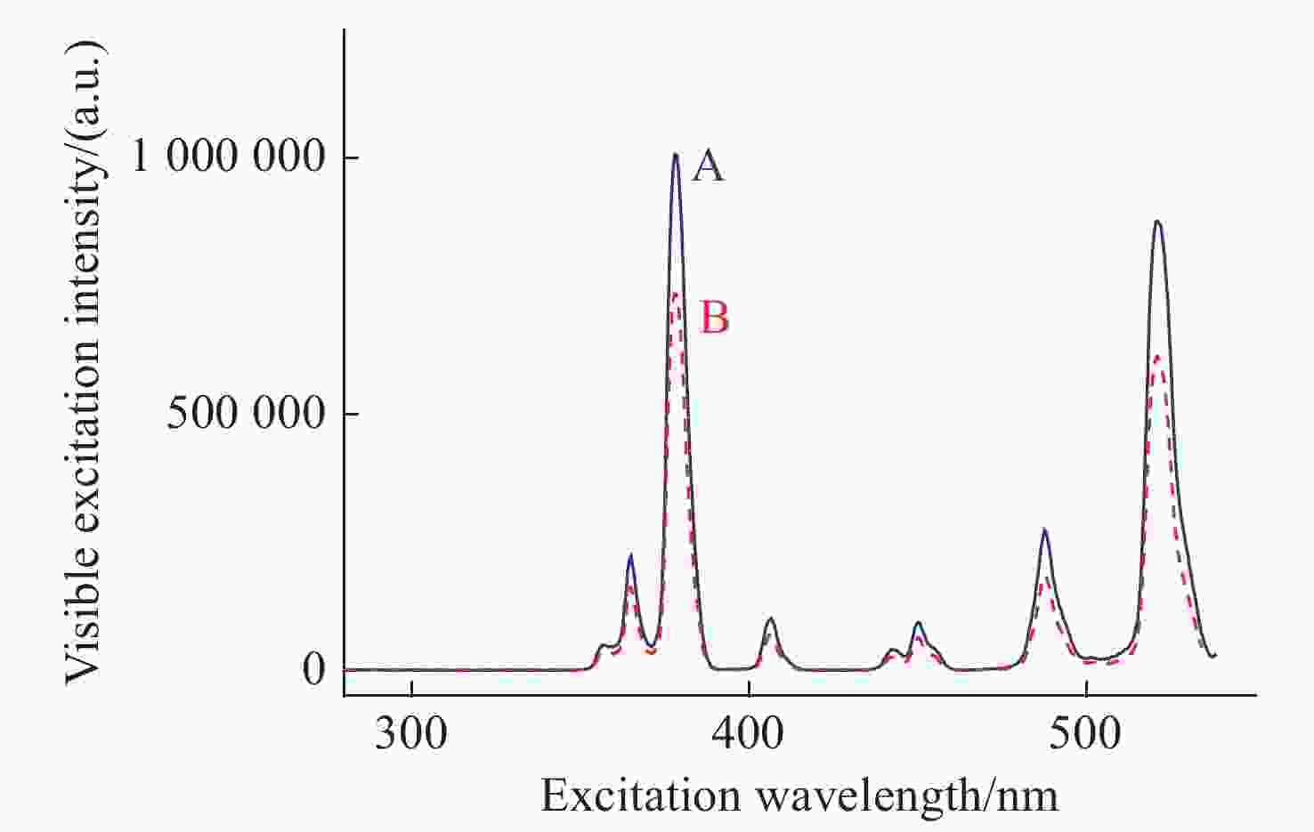
图 5 (A) Ag(1.6×10⁻⁶ mol /L)@SiO2(40 nm)@Er3+(0.5%): 铒碲发光玻璃样品(A蓝线)与(B) Er(0.5%): 铒碲发光玻璃样品(B红线)在280~538 nm波长范围内可见激发光谱(接收荧光波长为550 nm)
Figure 5. The visible excitation spectra of (A) Ag(1.6×10⁻⁶ mol/L)@SiO2(40 nm)@Er3+(0.5%):TeZnLa glass (blue line A) and (B) Er(0.5%):TeZnLa glass (red line B) from 280 nm to 538 nm when monitored at 550 nm
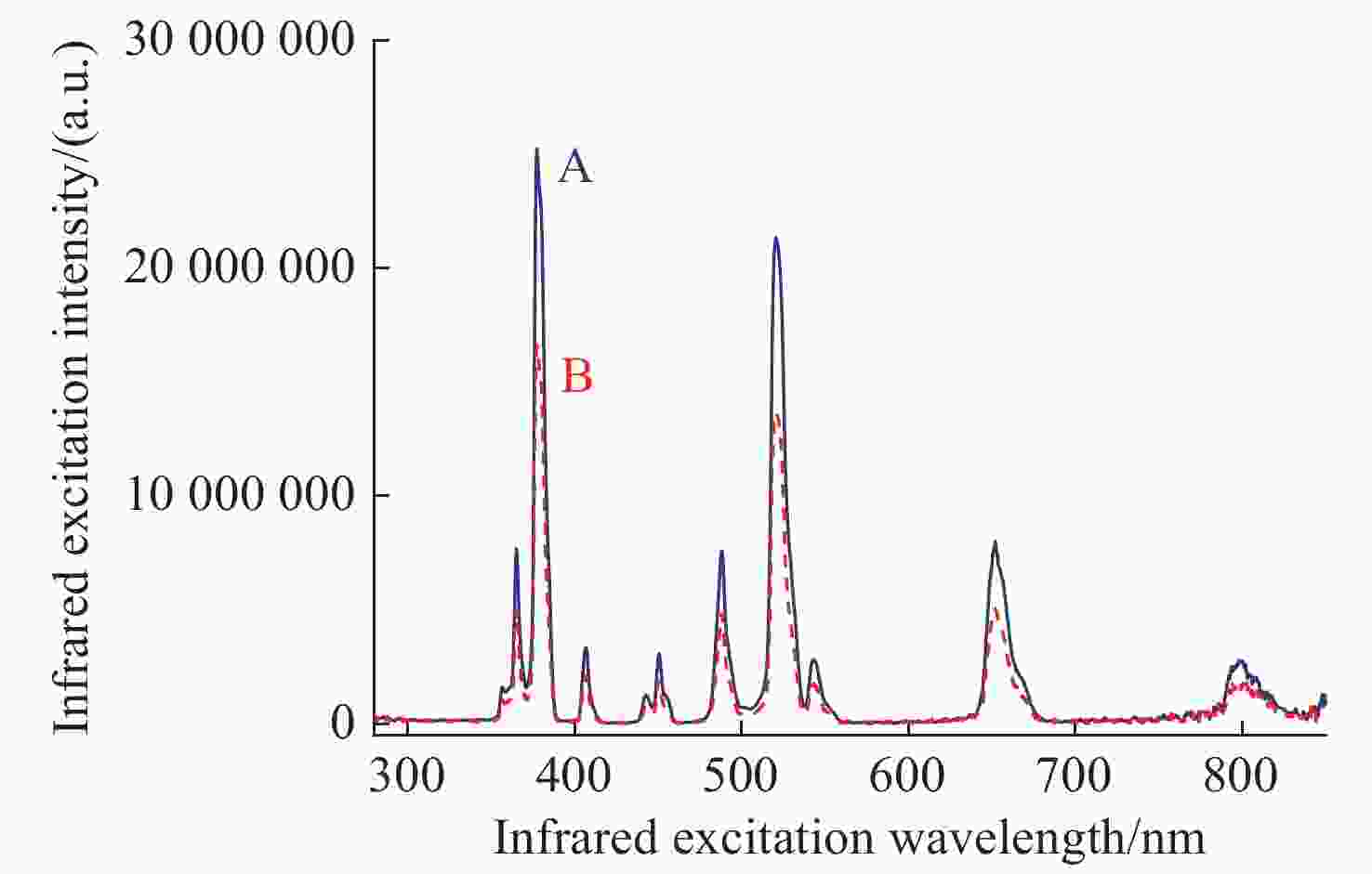
图 6 (A) Ag(1.6×10⁻⁶ mol/L)@SiO2(40 nm)@Er3+(0.5%):铒碲发光玻璃样品(A 蓝线)与(B) Er(0.5%):铒碲发光玻璃样品(B 红线)在280~850 nm波长范围内红外激发光谱(接收荧光波长为1531 mm)
Figure 6. The infrared excitation spectra of (A) Ag(1.6×10⁻⁶ mol/L)@SiO2(40 nm)@Er3+(0.5%):TeZnLa glass ((blue line A) and (B) Er(0.5%):TeZnLa glass (red line B) from 280 nm to 850 nm when monitored at 1531 nm
图 7 (A) Ag(1.6×10⁻⁶ mol /L)@SiO2(40 nm)@Er3+(0.5%): 铒碲发光玻璃样品(A 蓝线)与(B) Er3+(0.5%):铒碲发光玻璃样品(B 红线)在395 nm到718 nm波长范围内可见发光光谱(激发波长为378.0 nm)
Figure 7. The visible luminescence spectra of (A) Ag(1.6×10⁻⁶ mol/L)@SiO2(40 nm)@Er3+(0.5%):TeZnLa glass (blue line A) and (B) Er(0.5%):TeZnLa sample (red line B) from 395 nm to 718 nm when excited by 378.0 nm
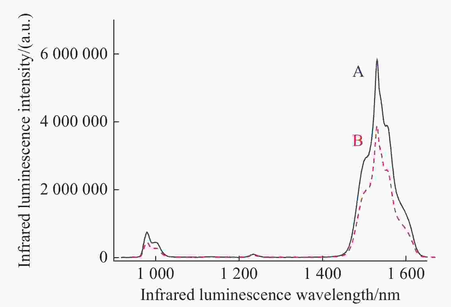
图 8 (A)Ag(1.6×10⁻⁶ mol/L)@SiO2(40 nm)@Er3+(0.5%): 铒碲发光玻璃样品(A 蓝线)与(B) Er3+(0.5%): 铒碲发光玻璃样品(B 红线)的918 nm到1680 nm波长范围的红外发光光谱(激发波长为378.0 nm)
Figure 8. The infrared luminescence spectra of (A) Ag(1.6×10⁻⁶ mol/L)@SiO2(40 nm)@Er3+(0.5%):TeZnLa glass (blue line A) and (B) Er(0.5%):TeZnLa glass (red line B) from 918 nm to 1680 nm when excited by 378.0 nm
图 9 (A) Ag(1.6×10⁻⁶ mol /L)@SiO2(40 nm)@Er3+(0.5%): 铒碲发光玻璃样品(A 蓝点)与(B) Er3+(0.5%): 铒碲发光玻璃样品(B 红点)在550.0 nm波长下的荧光寿命(激发波长为 378 nm)
Figure 9. The fluorescence lifetime of (A) Ag(1.6×10⁻⁶ mol /L)@SiO2(40 nm)@Er3+(0.5%):TeZnLa glass (blue dots A) and (B) Er(0.5%):TeZnLa glass (red dots B) at 550 nm luminescent wavelength were measured using a 378.0 nm pulsed xenon lamp as the pump source
表 1 样品A与样品B的可见光与红外光的发光强度与增强倍数
Table 1. The luminescence intensity and the enhancement factor of the visible and infrared luminescence of sample A and sample B
激发波长/nm 发光强度×105 增强数 样品A 样品B 546.0 nm 546.0 nm 546.0 nm 1531.0 nm 546.0 nm 1531.0 nm 378.0 2.261 58.22 1.501 38.80 150.6% 150.1% 406.5 0.222 − 0.143 − 155.2% − 520.5 2.090 51.49 1.376 33.96 151.9% 151.6% -
[1] XUE B, WANG D, ZHANG Y L, et al. Regulating the color output and simultaneously enhancing the intensity of upconversion nanoparticles via a dye sensitization strategy[J]. Journal of Materials Chemistry C, 2019, 7(28): 8607-8615. doi: 10.1039/C9TC02293G [2] LIN L, YU ZH P, WANG ZH ZH, et al. Plasmon-enhanced luminescence of Ag@SiO2/β-NaYF4: Tb3+ nanocomposites via absorption & emission matching[J]. Materials Chemistry and Physics, 2018, 220: 278-285. doi: 10.1016/j.matchemphys.2018.08.076 [3] ZHAO G Y, XU L ZH, MENG SH H, et al. Facile preparation of plasmon enhanced near-infrared photoluminescence of Er3+-doped Bi2O3-B2O3-SiO2 glass for optical fiber amplifier[J]. Journal of Luminescence, 2019, 206: 164-168. doi: 10.1016/j.jlumin.2018.10.026 [4] PARK W, LU D W, AHN S M. Plasmon enhancement of luminescence upconversion[J]. Chemical Society Reviews, 2015, 44(10): 2940-2962. doi: 10.1039/C5CS00050E [5] ZHAO J Y, CHENG Y Q, SHEN H M, et al. Light emission from plasmonic nanostructures enhanced with fluorescent nanodiamonds[J]. Scientific Reports, 2018, 8(1): 3605. doi: 10.1038/s41598-018-22019-z [6] CHEN G X, DING CH J, WU E, et al. Tip-enhanced upconversion luminescence in Yb3+-Er3+ codoped NaYF4 nanocrystals[J]. The Journal of Physical Chemistry C, 2015, 119(39): 22604-22610. doi: 10.1021/acs.jpcc.5b04387 [7] HE J J, ZHENG W, LIGMAJER F, et al. Plasmonic enhancement and polarization dependence of nonlinear upconversion emissions from single gold nanorod@SiO2@CaF2: Yb3+, Er3+ hybrid core-shell-satellite nanostructures[J]. Light:Science &Applications, 2017, 6(5): e16217. [8] WANG D, XUE B, TU L P, et al. Enhanced dye-sensitized up-conversion luminescence of neodymium-sensitized multi-shell nanostructures[J]. Chinese Optics, 2021, 14(2): 418-430. doi: 10.37188/CO.2020-0097 [9] YANG ZH, Ni W H, KOU X SH, et al. Incorporation of gold nanorods and their enhancement of fluorescence in mesostructured silica thin films[J]. The Journal of Physical Chemistry C, 2008, 112(48): 18895-18903. doi: 10.1021/jp8069699 [10] GEDDES C D, PARFENOV A, ROLL D, et al. Silver fractal-like structures for metal-enhanced fluorescence: enhanced fluorescence intensities and increased probe photostabilities[J]. Journal of Fluorescence, 2003, 13(3): 267-276. doi: 10.1023/A:1025046101335 [11] WANG Q R, ZHANG J, SANG X, et al. Enhanced luminescence and prolonged lifetime of Eu-PMMA films based on Au@SiO2 plasmonic hetero-nanorods[J]. Journal of Luminescence, 2018, 204: 284-288. doi: 10.1016/j.jlumin.2018.08.033 [12] XU W, LEE T K, MOON B S, et al. Broadband plasmonic antenna enhanced upconversion and its application in flexible fingerprint identification[J]. Advanced Optical Materials, 2018, 6(6): 1701119. doi: 10.1002/adom.201701119 [13] RAJESH D, DOUSTI M R, AMJAD R J, et al. Enhancement of down- and upconversion intensities in Er3+/Yb3+ co-doped oxyfluoro tellurite glasses induced by Ag species and nanoparticles[J]. Journal of Luminescence, 2017, 192: 250-255. doi: 10.1016/j.jlumin.2017.06.059 [14] DAS A, MAO CH CH, CHO S, et al. Over 1000-fold enhancement of upconversion luminescence using water-dispersible metal-insulator-metal nanostructures[J]. Nature Communications, 2018, 9(1): 4828. doi: 10.1038/s41467-018-07284-w [15] FARES H, ELHOUICHET H, GELLOZ B, et al. Silver nanoparticles enhanced luminescence properties of Er3+ doped tellurite glasses: effect of heat treatment[J]. Journal of Applied Physics, 2014, 116(12): 123504. doi: 10.1063/1.4896363 [16] 徐光宪. 稀土[M]. 2版. 北京: 冶金工业出版社, 1995.XU G X. Rare Earth[M]. 2nd ed. Beijing: Metallurgical Industry Press, 1995. (in Chinese) [17] 郭光灿, 金怀诚, 谢建平. 光学原子物理[M]. 合肥: 中国科学技术大学出版社, 1990.GUO G C, JIN H CH, XIE J P. Optical Atomic Physics[M]. Hefei: China University of Science and Technology Press, 1990. (in Chinese) [18] 王永生, 张雪强, 张光寅, 等. BaFCl: Eu2+中F色心的浓度和光激励截面与温度和紫外线的辐照波长的关系[J]. 发光学报,1996,17(1):6-11. doi: 10.3321/j.issn:1000-7032.1996.01.002WANG Y SH, ZHANG X Q, ZHANG G Y, et al. The dependence of density and photostimulable cross section of F color centers in BaFCl: Eu2+ phosphors on temperature and UV-irradiation wavelength[J]. Chinese Journal of Luminescence, 1996, 17(1): 6-11. (in Chinese) doi: 10.3321/j.issn:1000-7032.1996.01.002 [19] 彭皓, 杨方, 杜慧, 等. 基于Er3+掺杂上转换纳米粒子的生物成像研究进展[J]. 分析化学,2021,49(7):1106-1120.PENG H, YANG F, DU H, et al. Advances of Er3+ doped upconversion nanoparticles for biological imaging[J]. Chinese Journal of Analytical Chemistry, 2021, 49(7): 1106-1120. (in Chinese) [20] 安西涛, 王月, 牟佳佳, 等. 超薄金壳包覆NaYF4: Yb, Er@SiO2纳米结构的可控合成与表面增强上转换荧光[J]. 发光学报,2018,39(11):1505-1512. doi: 10.3788/fgxb20183911.1505AN X T, WANG Y, MU J J, et al. Controllable synthesis and surface-enhanced upconversion luminescence of ultra-thin gold shell coated NaYF4: Yb, Er@SiO2 nanostructures[J]. Chinese Journal of Luminescence, 2018, 39(11): 1505-1512. (in Chinese) doi: 10.3788/fgxb20183911.1505 [21] 胡家乐, 薛冬峰. 稀土离子特性与稀土功能材料研究进展[J]. 应用化学,2020,37(3):245-255. doi: 10.11944/j.issn.1000-0518.2020.03.190350HU J L, XUE D F. Research progress on the characteristics of rare earth ions and rare earth functional materials[J]. Chinese Journal of Applied Chemistry, 2020, 37(3): 245-255. (in Chinese) doi: 10.11944/j.issn.1000-0518.2020.03.190350 [22] 李子娟, 安雪, 牛昊, 等. 高温溶剂热分解法合成NaYF4: Yb3+, Er3+纳米粒子及其光谱特性[J]. 发光学报,2020,41(9):1128-1136. doi: 10.37188/fgxb20204109.1128LI Z J, AN X, NIU H, et al. Synthesis and spectral properties of NaYF4: Yb3+, Er3+ nanoparticles via thermolysis method[J]. Chinese Journal of Luminescence, 2020, 41(9): 1128-1136. (in Chinese) doi: 10.37188/fgxb20204109.1128 [23] 赵兵洁, 赵金宝, 齐小花, 等. 基于BHHCT-Eu3+@SiO2荧光稀土二氧化硅纳米颗粒的免疫层析试纸条检测卡那霉素[J]. 分析化学,2017,45(10):1467-1474. doi: 10.11895/j.issn.0253-3820.170015ZHAO B J, ZHAO J B, QI X H, et al. Development of immunochromatographic strips based on covalently conjugated BHHCT-Eu3+@SiO2 for rapid and quantitative detection of kanamycin[J]. Chinese Journal of Analytical Chemistry, 2017, 45(10): 1467-1474. (in Chinese) doi: 10.11895/j.issn.0253-3820.170015 -


 下载:
下载:

