


Longitudinal chromatic aberration compensation method for dual-wavelength retinal imaging adaptive optics systems
doi: 10.37188/CO.EN.2021-0009
-
摘要: 双波长视网膜成像自适应光学系统非常适用于视网膜微血管的高对比度和高分辨率成像。本文重点研究了双波长自适应系统的轴向色差补偿问题。首先对轴向色差进行了测量,对实测波前进行了分析,并给出任意波前轴向色差补偿法。自适应校正实验结果显示,色差补偿后,波前均方根误差减小到0.16 λ(λ=589 nm),视网膜微血管分辨率提高到6 μm。这项工作可用于视网膜成像的临床应用。Abstract: Dual-wavelength retinal imaging adaptive optics systems are suitable for high contrast and resolution imaging of retinal capillaries. The compensation of the Longitudinal Chromatic Aberrations (LCAs) in dual-wavelength adaptive systems is researched. The LCA is measured, the measured wavefronts are analyzed, and the arbitrary wavefront LCA compensation method is given. An adaptive correction experiment is carried out and the experimental results indicate that the root mean square error of the wavefront is reduced to 0.16 λ (λ=589 nm) and the retinal capillary resolution is improved to 6 μm. This work may be used for the clinical applications of retinal imaging.
-
Key words:
- adaptive optics /
- retinal imaging /
- longitudinal chromatic aberration /
- dual-wavelength
-
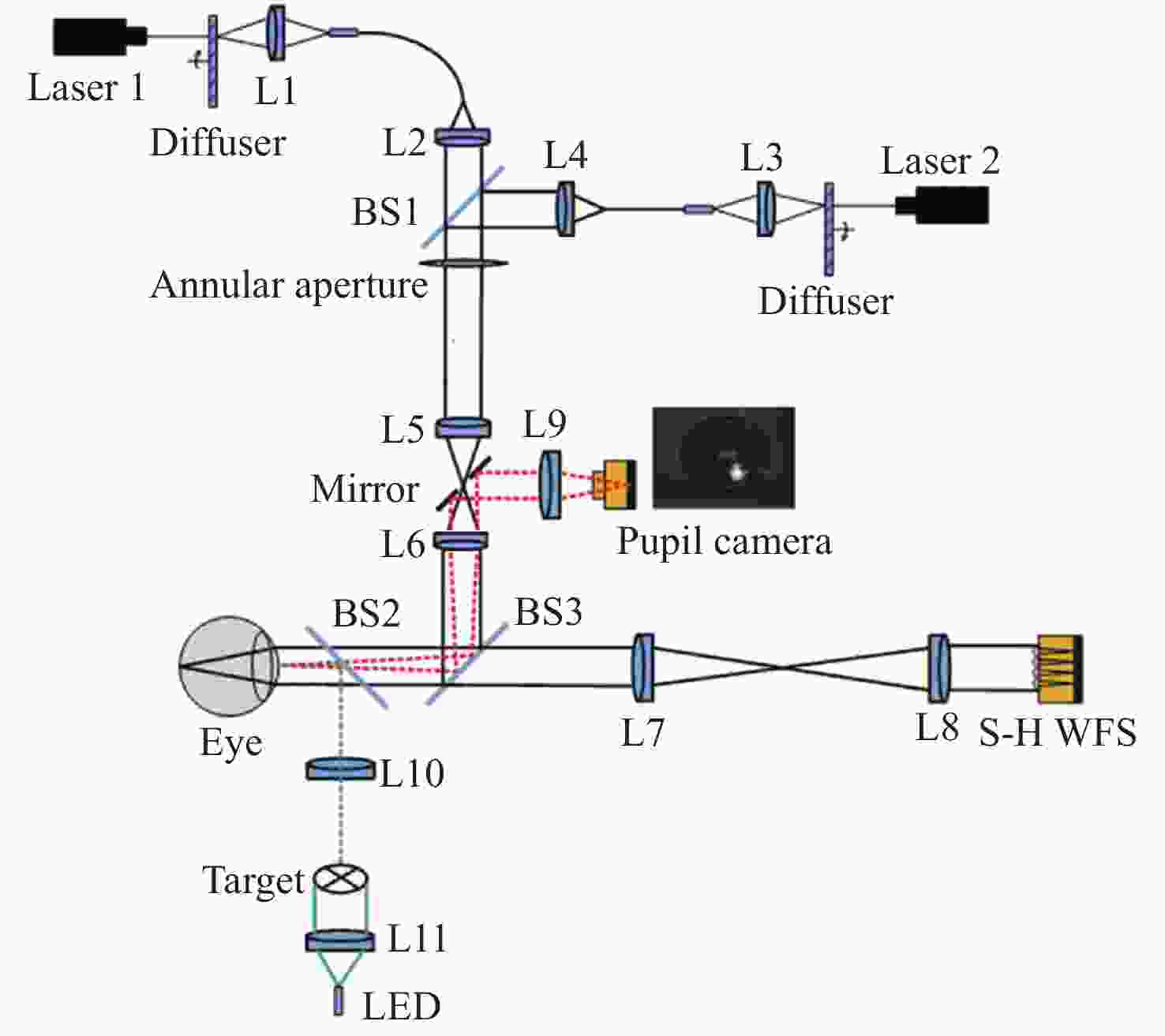
Figure 1. Illustration of the optical setup for LCA measurement: two lasers are used with the wavelength of 589 nm and 808 nm

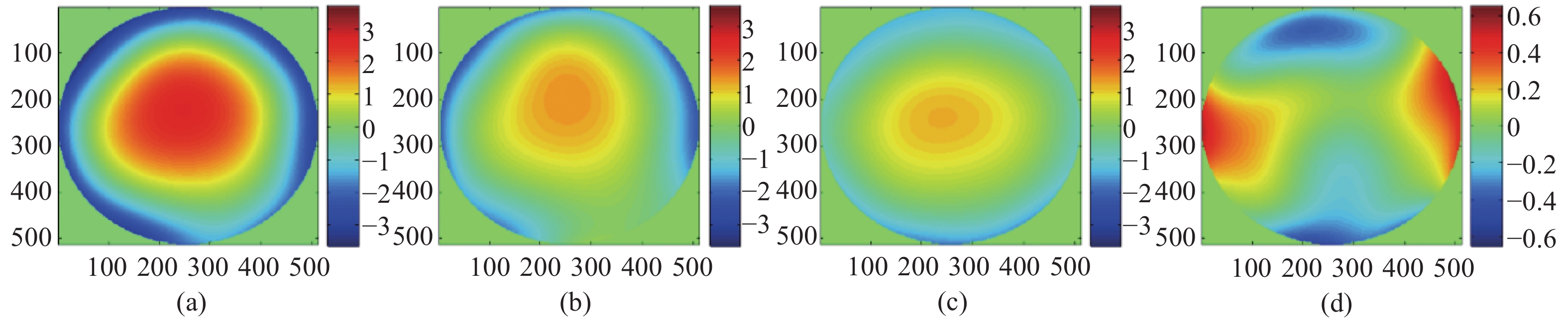
Figure 3. Measured wavefronts of subject A. (a) Wavelength of 589 nm; (b) wavelength of 808 nm; (c) LCA; (d) LCA without defocus
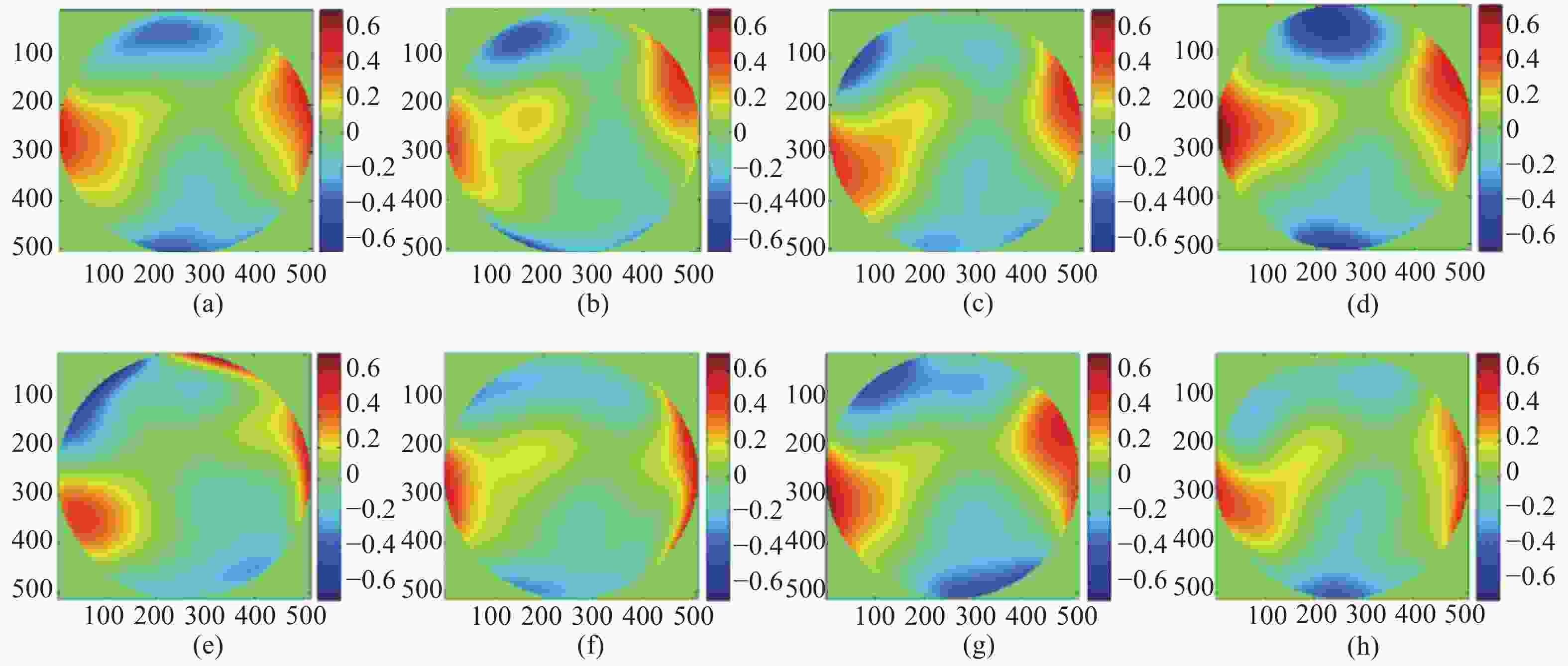
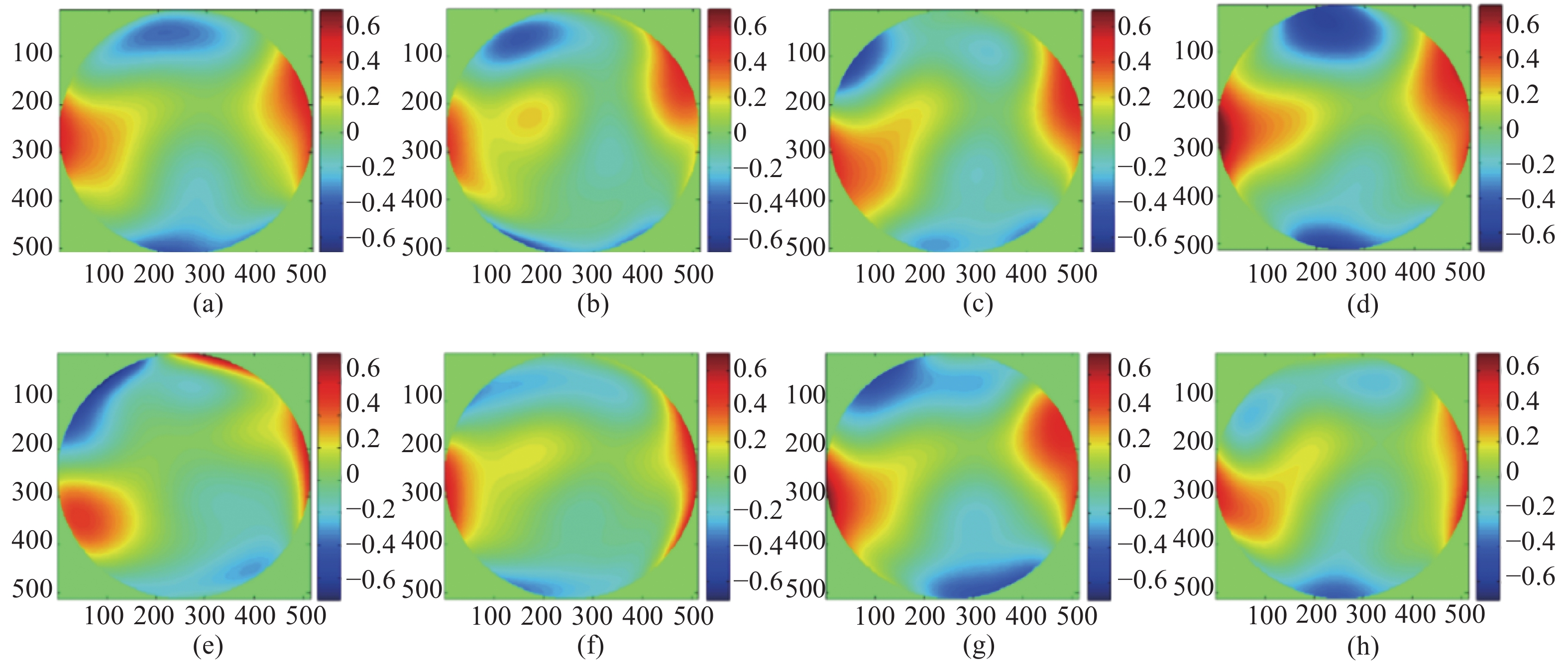
Figure 5. Wavefronts of LCA at different times for subject A. (a) Start; (b) 5 minutes later; (c) 1 hour later; (d) 10 hours later; (e) 15 hours later; (f) 24 hours later; (g) 30 hours later; (h) 36 hours later
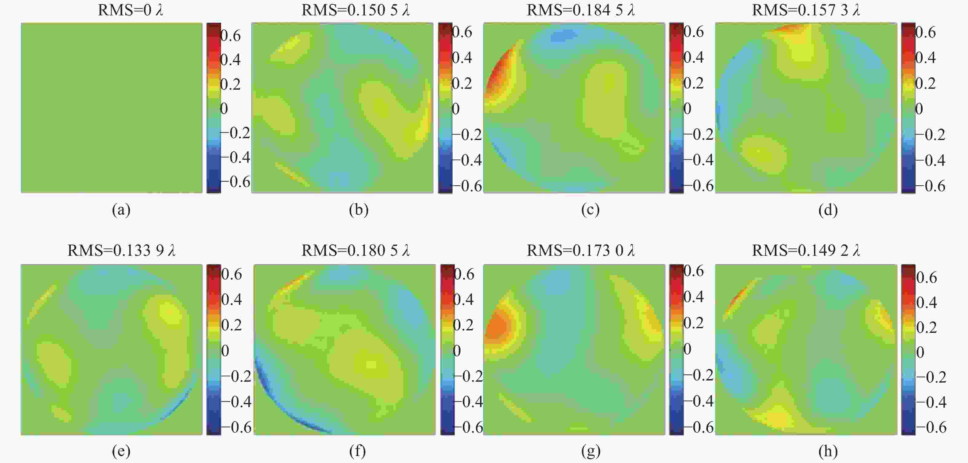
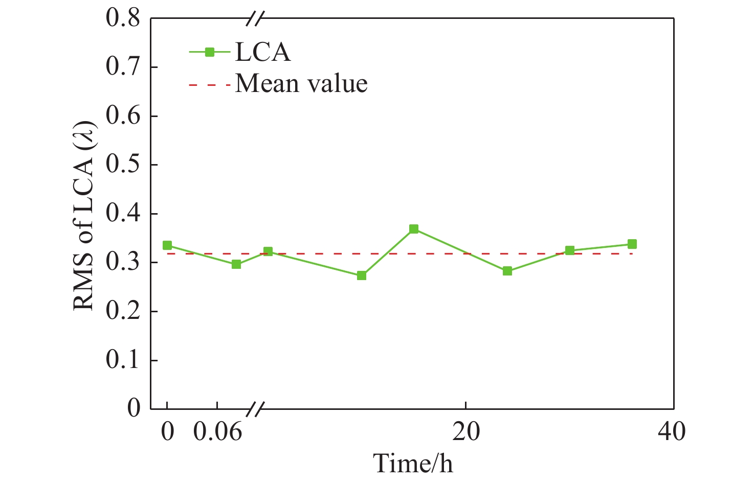
Figure 7. Calculated compensation of LCA for subject A at different times while the first wavefront of Fig. 5 is chosen as the arbitrary wavefront. The mean value of LCA is 0.16 λ with the standard deviation of 0.017. (a) Start; (b) 5 minutes later; (c) 1 hour later; (d) 10 hours later; (e) 15 hours later; (f) 24 hours later; (g) 30 hours later; (h) 36 hours later
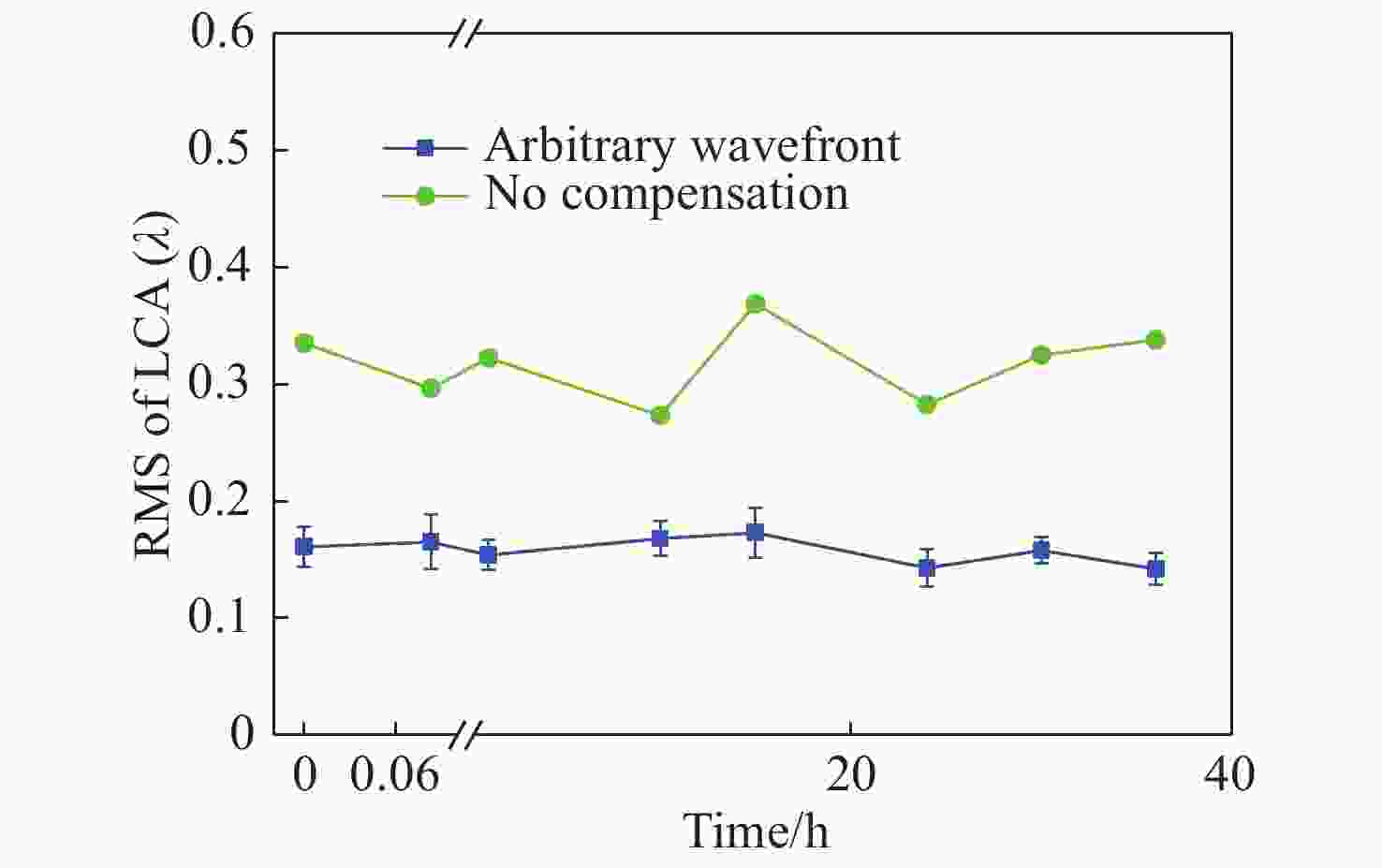
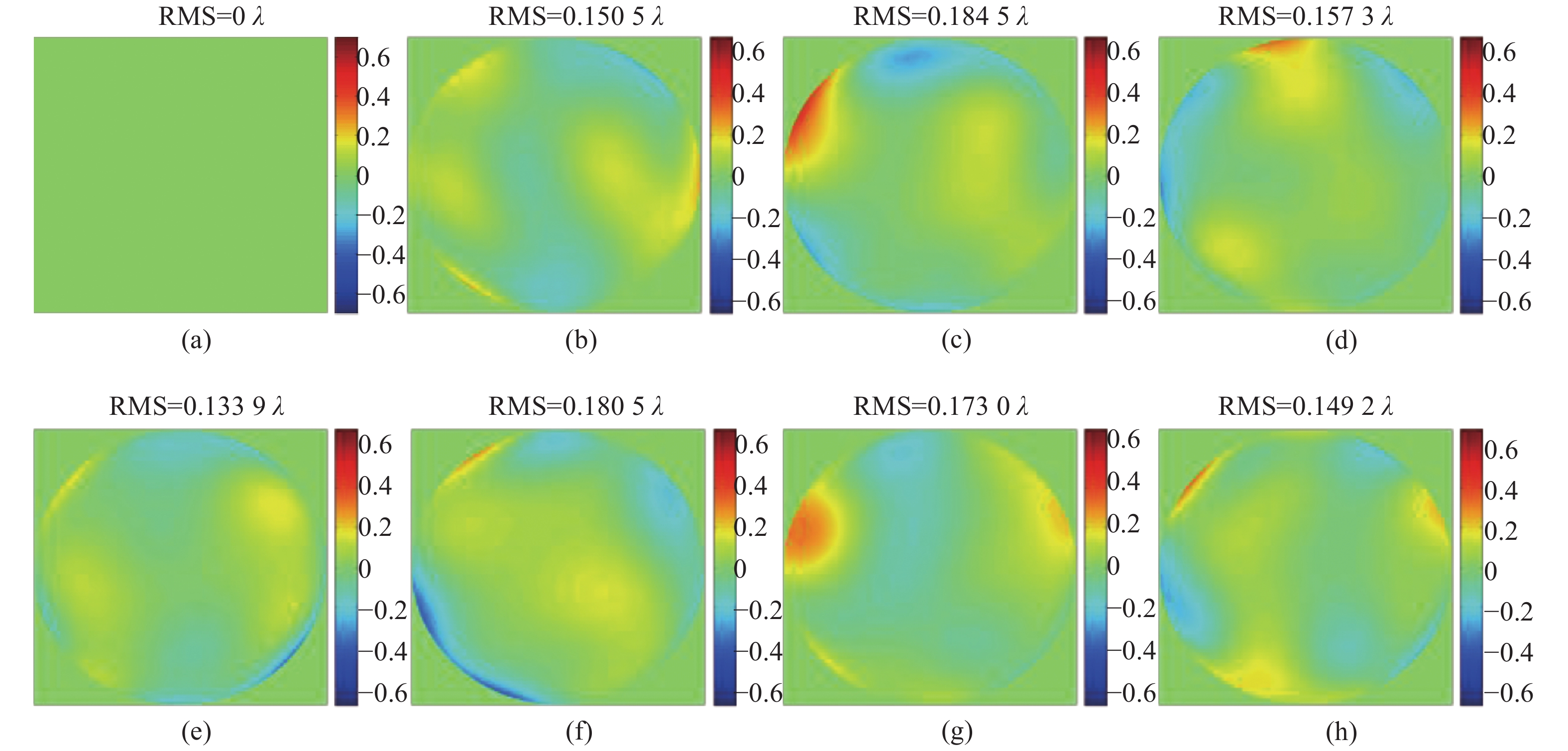
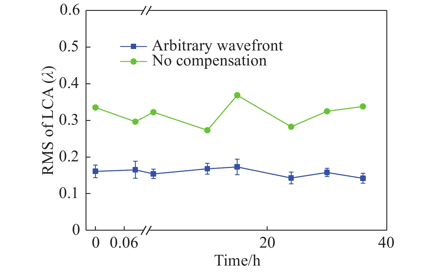
Figure 8. Compensation of LCA for subject A at different times. The mean value of the arbitrary wavefront is 0.158±0.011 λ.
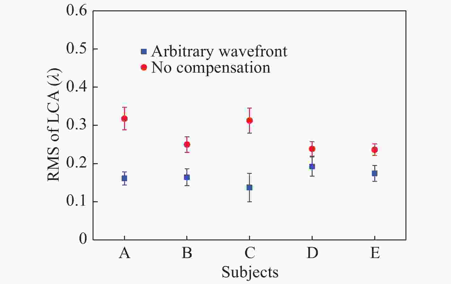
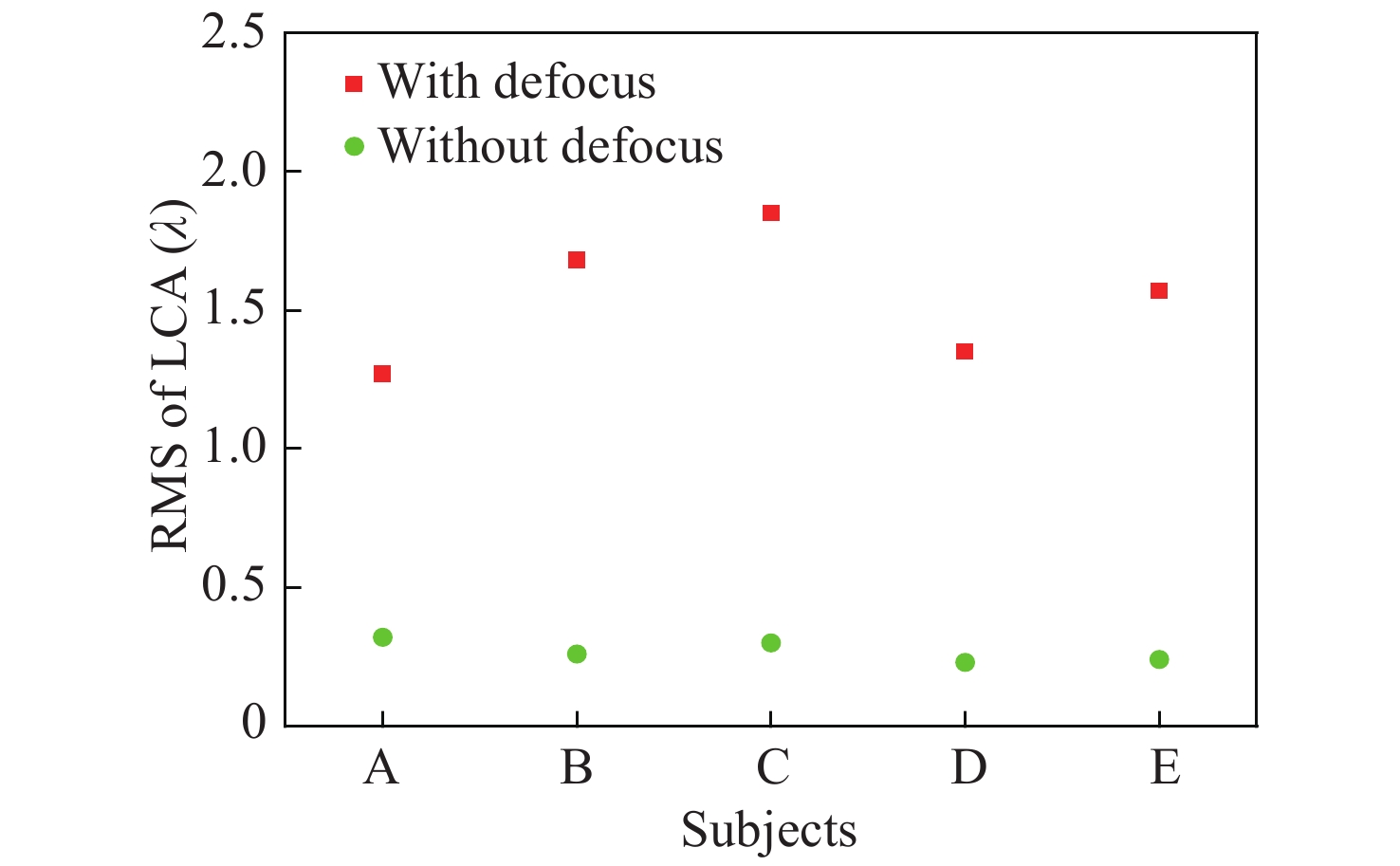
Figure 9. Compensation of LCA for different subjects. The mean values of the arbitrary wavefront is 0.166±0.017 λ.
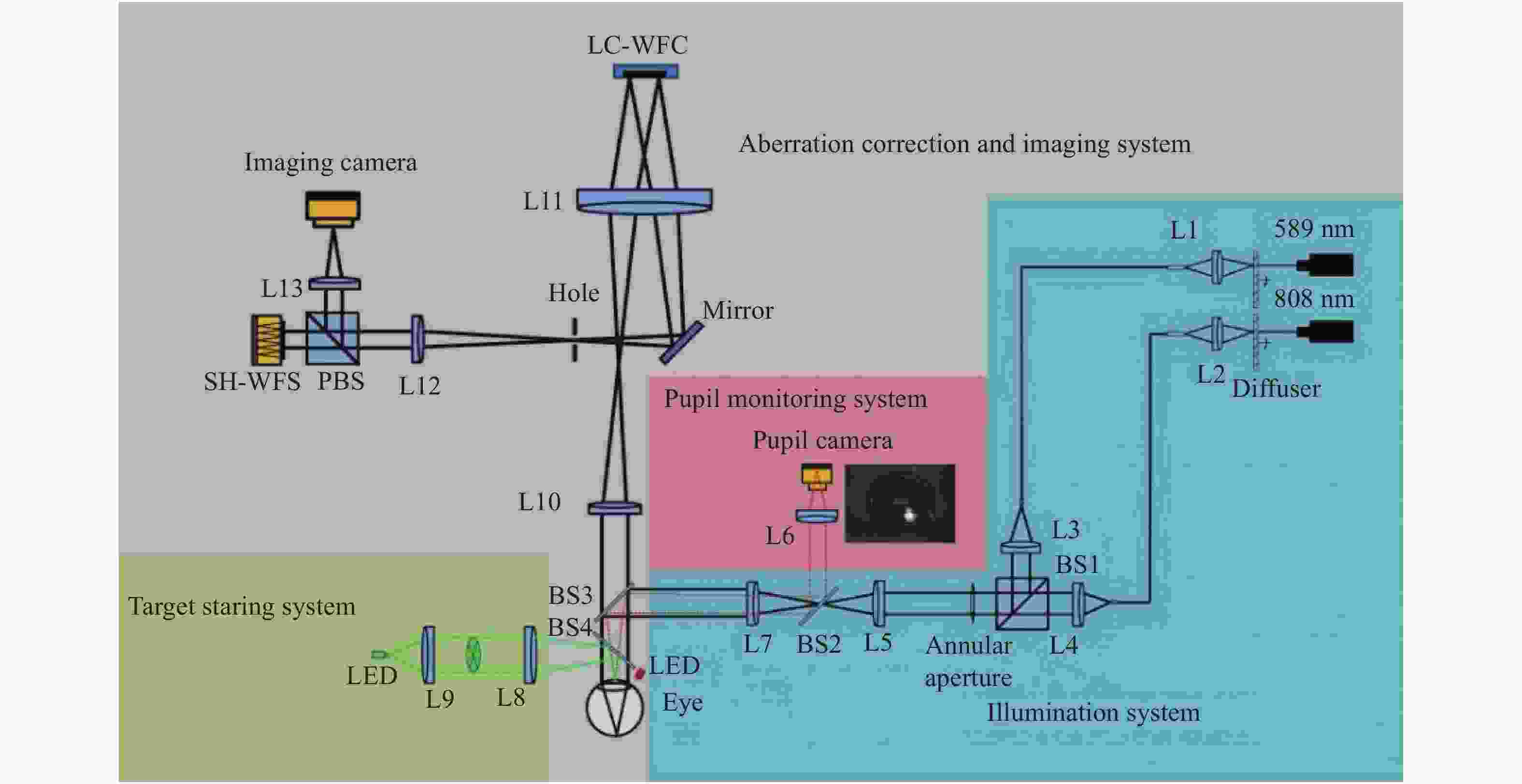
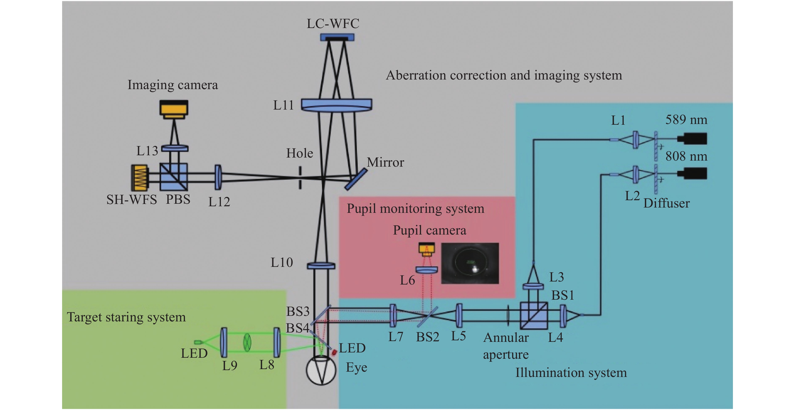
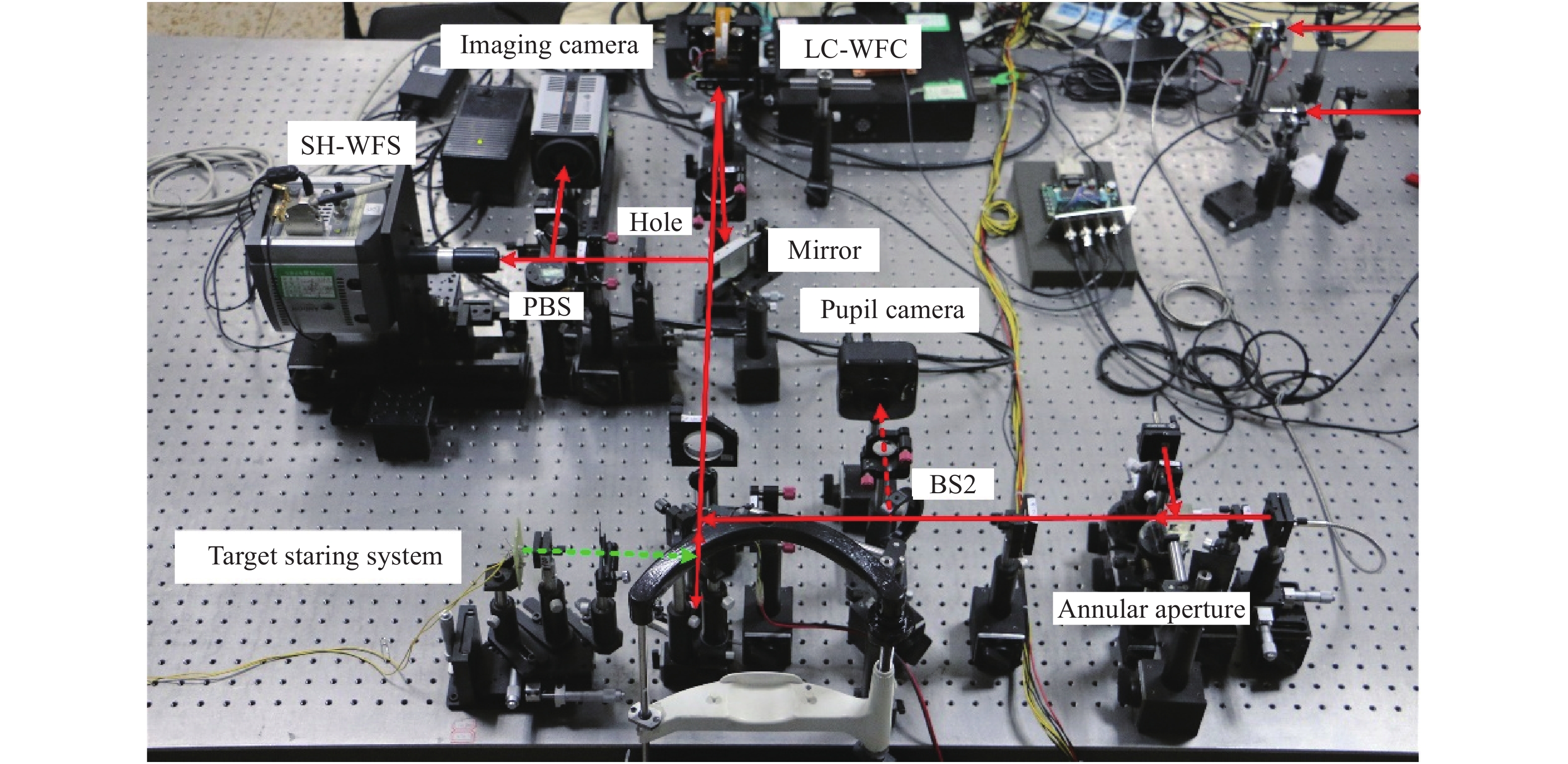
Figure 10. Optical layout for the retinal imaging AOS: L1-L13, Lens 1- Lens 13; PBS, polarizing beam splitter; BS, beam splitter; an 808 nm laser is used for wavefront detection, tracing and positioning the capillary; a 589 nm laser is used for high contrast imaging of the capillary; the collimated beam comes from the illumination system and is reflected into the eye, and then reflected again out from the eye by the retina; this reflected light is detected and corrected by the adaptive optics system and imaged with an imaging camera; the pupil position is observed by the pupil monitoring system and the eye is fixed with the target staring system

Figure 12. Experiment results of adaptive correction and LCA compensation for subjects A and C. (a) Measured aberration at 808 nm for subject A; (b) wavefront of LCA for subject A; (c) image of retinal capillary without LCA compensation for subject A; (d) image of retinal capillary after LCA compensation for subject A; (e) measured aberration at 808 nm for subject C; (f) wavefront of LCA for subject C; (g) image of retinal capillary without LCA compensation for subject C; (h) image of retinal capillary after LCA compensation for subject C
-
[1] LIANG J ZH, WILLIAMS D R, MILLER D T. Supernormal vision and high-resolution retinal imaging through adaptive optics[J]. Journal of the Optical Society of America A, 1997, 14(11): 2884-2892. doi: 10.1364/JOSAA.14.002884 [2] ROORDA A, WILLIAMS D R. The arrangement of the three cone classes in the living human eye[J]. Nature, 1999, 397(6719): 520-522. doi: 10.1038/17383 [3] JONNAL R S, RHA J, ZHANG Y, et al. In vivo functional imaging of human cone photoreceptors[J]. Optics Express, 2007, 15(24): 16141-16160. doi: 10.1364/OE.15.016141 [4] LI K Y, ROORDA A. Automated identification of cone photoreceptors in adaptive optics retinal images[J]. Journal of the Optical Society of America A, 2007, 24(5): 1358-1363. doi: 10.1364/JOSAA.24.001358 [5] DUBRA A, SULAI Y, NORRIS J L, et al. Noninvasive imaging of the human rod photoreceptor mosaic using a confocal adaptive optics scanning ophthalmoscope[J]. Biomedical Optics Express, 2011, 2(7): 1864-1876. doi: 10.1364/BOE.2.001864 [6] CHUI T Y P, VANNASDALE D A, BURNS S A. The use of forward scatter to improve retinal vascular imaging with an adaptive optics scanning laser ophthalmoscope[J]. Biomedical Optics Express, 2012, 3(10): 2537-2549. doi: 10.1364/BOE.3.002537 [7] ZAYIT-SOUDRY S, DUNCAN J L, SYED R, et al. Cone structure imaged with adaptive optics scanning laser ophthalmoscopy in eyes with nonneovascular age-related macular degeneration[J]. Investigative Ophthalmology &Visual Science, 2013, 54(12): 7498-7509. [8] QUERQUES G, KAMAMI-LEVY C, GEORGES A, et al. Appearance of regressing drusen on adaptive optics in age-related macular degeneration[J]. Ophthalmology, 2014, 121(2): 611-612. doi: 10.1016/j.ophtha.2013.10.006 [9] PAQUES M, BROLLY A, BENESTY J, et al. Venous nicking without arteriovenous contact: the role of the arteriolar microenvironment in arteriovenous nickings[J]. JAMA Ophthalmology, 2015, 133(8): 947-950. doi: 10.1001/jamaophthalmol.2015.1132 [10] MARTIN J A, ROORDA A. Direct and noninvasive assessment of parafoveal capillary leukocyte velocity[J]. Ophthalmology, 2005, 112(12): 2219-2224. doi: 10.1016/j.ophtha.2005.06.033 [11] HAN G Q. High contrast imaging study of retinal capillaries in human eyes[D]. Beijing: Changchun Institute of Optics, Fine Mechanics and Physics University of Chinse Academy of Sciences, 2018: 28-30. (in Chinese) [12] FABER D J, AALDERS M C G, MIK E G, et al. Oxygen saturation-dependent absorption and scattering of blood[J]. Physical Review Letters, 2004, 93(2): 028102. doi: 10.1103/PhysRevLett.93.028102 [13] REINHOLZ F, ASHMAN R A, EIKELBOOM R H. Simultaneous three wavelength imaging with a scanning laser ophthalmoscope[J]. Cytometry Part A, 1999, 37(3): 165-170. doi: 10.1002/(SICI)1097-0320(19991101)37:3<165::AID-CYTO1>3.0.CO;2-A [14] GRIEVE K, TIRUVEEDHULA P, ZHANG Y H, et al. Multi-wavelength imaging with the adaptive optics scanning laser ophthalmoscope[J]. Optics Express, 2006, 14(25): 12230-12242. doi: 10.1364/OE.14.012230 [15] NEWTON S I. Opticks, or, A Treatise of the Reflections, Refractions, in Flections & Colours of Light[M]. 4th ed. London: G. Bell & Sons, Ltd. , 1931. [16] WALD G, GRIFFIN D R. The change in refractive power of the human eye in dim and bright light[J]. Journal of the Optical Society of America, 1947, 37(5): 321-336. doi: 10.1364/JOSA.37.000321 [17] THIBOS L N, BRADLEY A, STILL D L, et al. Theory and measurement of ocular chromatic aberration[J]. Vision Research, 1990, 30(1): 33-49. doi: 10.1016/0042-6989(90)90126-6 [18] FERNÁNDEZ E J, ARTAL P. Ocular aberrations up to the infrared range: from 632.8 to 1070 nm[J]. Optics Express, 2008, 16(26): 21199-21208. doi: 10.1364/OE.16.021199 [19] RUCKER F J, OSORIO D. The effects of longitudinal chromatic aberration and a shift in the peak of the middle-wavelength sensitive cone fundamental on cone contrast[J]. Vision Research, 2008, 48(19): 1929-1939. doi: 10.1016/j.visres.2008.06.021 [20] NAKAJIMA M, HIRAOKA T, HIROHARA Y, et al. Verification of the lack of correlation between age and longitudinal chromatic aberrations of the human eye from the visible to the infrared[J]. Biomedical Optics Express, 2015, 6(7): 2676-2694. doi: 10.1364/BOE.6.002676 [21] VINAS M, DORRONSORO C, CORTES D, et al. Longitudinal chromatic aberration of the human eye in the visible and near infrared from wavefront sensing, double-pass and psychophysics[J]. Biomedical Optics Express, 2015, 6(3): 948-962. doi: 10.1364/BOE.6.000948 [22] VINAS M, DORRONSORO C, GARZÓN N, et al. In vivo subjective and objective longitudinal chromatic aberration after bilateral implantation of the same design of hydrophobic and hydrophilic intraocular lenses[J]. Journal of Cataract &Refractive Surgery, 2015, 41(10): 2115-2124. [23] CHONG S P, ZHANG T W, KHO A, et al. Ultrahigh resolution retinal imaging by visible light OCT with longitudinal achromatization[J]. Biomedical Optics Express, 2018, 9(4): 1477-1491. doi: 10.1364/BOE.9.001477 [24] ZAWADZKI R J, CENSE B, ZHANG Y, et al. Ultrahigh-resolution optical coherence tomography with monochromatic and chromatic aberration correction[J]. Optics Express, 2008, 16(11): 8126-8143. doi: 10.1364/OE.16.008126 [25] DUBRA A, SULAI Y. Reflective afocal broadband adaptive optics scanning ophthalmoscope[J]. Biomedical Optics Express, 2011, 2(6): 1757-1768. doi: 10.1364/BOE.2.001757 [26] JIANG X Y, KUCHENBECKER J A, TOUCH P, et al. Measuring and compensating for ocular longitudinal chromatic aberration[J]. Optica, 2019, 6(8): 981-990. doi: 10.1364/OPTICA.6.000981 [27] ZHENG X L, LIU R X, XIA M L, et al. Temporal properties study of ocular wave aberrations with high frequency sampling[J]. Chinese Optics, 2014, 34(7): 0733001. [28] MORGAN J I W, HUNTER J J, MASELLA B, et al. Light-induced retinal changes observed with high-resolution autofluorescence imaging of the retinal pigment epithelium[J]. Investigative Ophthalmology &Visual Science, 2008, 49(8): 3715-3729. [29] DELORI F C, WEBB R H, SLINEY D H. Maximum permissible exposures for ocular safety (ANSI 2000), with emphasis on ophthalmic devices[J]. Journal of the Optical Society of America A, 2007, 24(5): 1250-1265. doi: 10.1364/JOSAA.24.001250 [30] Laser Institute of America. ANSI Z136.1-2014 American national standard for safe use of lasers[S]. Orlando: American National Standards Institute, 2007: 22-29. [31] LI CH, XIA M L, MU Q Q, et al. High-precision open-loop adaptive optics system based on LC-SLM[J]. Optics Express, 2009, 17(13): 10774-10781. doi: 10.1364/OE.17.010774 [32] YANG L B, HU L F, LI D Y, et al. Determining the imaging plane of a retinal capillary layer in adaptive optical imaging[J]. Chinese Physics B, 2016, 25(9): 094219. doi: 10.1088/1674-1056/25/9/094219 [33] BURNS S A, ELSNER A E, CHUI T Y, et al. In vivo adaptive optics microvascular imaging in diabetic patients without clinically severe diabetic retinopathy[J]. Biomedical Optics Express, 2014, 5(3): 961-974. doi: 10.1364/BOE.5.000961 -

 下载:
下载:


计量
- 文章访问数: 1631
- HTML全文浏览量: 972
- PDF下载量: 314
- 被引次数: 0
