


Detection system of multilayer coating microstructure defects based on differential interference contrast confocal microscopy
-
摘要: 多层膜极紫外光刻掩模"白板"缺陷是制约下一代光刻技术发展的瓶颈之一,为提高对掩模"白板"上的膜层微结构缺陷的分辨能力,提出了一种微分干涉差共焦显微探测系统方案。基于标量衍射理论,计算了系统横向和轴向分辨率。利用MATLAB建模仿真,在数值孔径为0.65、工作波长为405 nm时,分析比较了微分干涉差共焦显微系统、传统显微系统和共焦显微系统的分辨率。结果表明微分干涉差共焦显微系统具有230 nm的横向分辨率和25 nm轴向台阶高度差的分辨能力(对应划痕等缺陷形式)。此外,仿真和分析了实际应用中探测器尺寸、样品轴向偏移等的影响,模拟分析了膜层微结构缺陷的探测,结果表明本系统可以探测200 nm宽、10 nm高的微结构缺陷,较另外两种系统有更好的探测能力。Abstract: Defects in the multilayer extreme ultraviolet lithography(EUV) mask "whiteboard" have become an important issue to restrict the development of next-generation lithography. A detection system based on differential interference contrast(DIC) confocal microscopy is proposed in order to improve the ability of distinguishing the microstructure defects on the lithography mask "whiteboard". Based on the scalar diffraction theory, the lateral and axial resolution of the system are calculated. In the condition that numerical aperture of 0.65 and working wavelength of 405 nm, resolutions of DIC confocal microscopy, traditional microscopy and the confocal microscopy system are compared and analyzed using MATLAB. The results show that the DIC confocal microscopy has the ability of lateral resolution of 230 nm and resolution of axial step height difference of 25 nm(corresponding to defects such as scratches). In addition, the effect of factors such as the size of detector and the axial deviation of the sample are also simulated and analyzed. The experimental results show that the proposed system can detect multilayer coating microstructure defects with a width of 200 nm and a height of 10 nm, which has better detection ability than the other two systems.
-

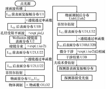
图 2 反射式DIC共焦系统等效光路图
Figure 2. Equivalent optical path of reflecting DIC confocal system

图 3 共焦和DIC共焦系统归一化光强分布曲线
Figure 3. Uniformization intensity curves of confocal and DIC confocal system

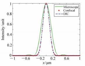
图 6 3种系统的横向归一化光强分布曲线对比
Figure 6. Comparison of uniformization lateral intensity distribution curves of 3 systems

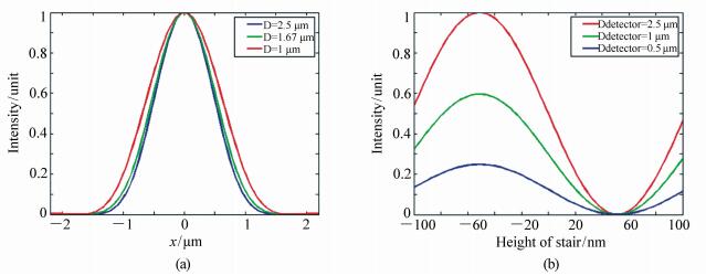
图 8 (a) 探测器直径分别为2.5 μm, 1.67 μm, 1 μm时共焦系统的轴向光强响应曲线; (b)探测器直径为2.5 μm, 1 μm, 0.5 μm时的DIC共焦系统的台阶光强响应曲线
Figure 8. (a)Intensity curves of confocal system with detector diameter of 2.5 μm, 1.67 μm, 1 μm; (b)Intensity curves of DIC confocal systems with detector diameter of 2.5 μm, 1 μm, 0.5 μm

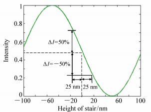
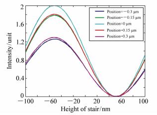
图 9 不同轴向偏移时DIC共焦系统的台阶响应
Figure 9. Stair response curves of DIC confocal system under different z-axial offsets
表 1 共焦和DIC共焦系统仿真值与理论值拟合结果
Table 1. Comparison between simulation and theoretical result of two systems
系统名称 横向分布相关系数 轴向分布相关系数 共焦系统 1 0.997 0 DIC共焦系统 1 0.996 3  下载: 导出CSV
表 2 3个系统横向分辨率比较
Table 2. Comparison of lateral resolutions among the 3 systems
(μm) 系统名称 半高全宽(FWHM) 传统显微镜 0.32 共焦系统 0.23 DIC共焦系统 0.23
下载: 导出CSV
表 3 共焦系统与DIC共焦系统镜面反射拟合结果
Table 3. Specular reflection fitting result of confocal and DIC confocal system
系统名称 相关系数R 共焦系统 0.997 2 DIC共焦系统 0.996 8
下载: 导出CSV
样品轴向偏移/μm 相关系数R -0.3 0.998 5 -0.15 0.999 1 0 0.999 3 0.15 0.999 1 0.3 0.998 7
下载: 导出CSV
表 5 不同尺寸缺陷对应的归一化探测光强
Table 5. Uniformization intensity of micro-defects with different sizes
z/nm d/nm 200 240 280 -20 1.141 0 1.254 2 1.280 5 -10 1.066 8 1.125 0 1.138 1 0 1.000 0 1.000 0 1.000 0 10 0.942 2 0.880 0 0.867 6 20 0.895 0 0.766 5 0.742 5
下载: 导出CSV
-
[1] 刘晓雷, 李思坤, 王向朝.极紫外光刻含缺陷多层膜衍射谱仿真简化模型[J].光学学报, 2014, 34(9):40-46. http://www.opticsjournal.net/abstract.htm?aid=OJ0815000014jPmSpVLIU X L, LI S K, WANG X ZH. Simplified model for defective multilayer diffraction spectrum simulation in extreme ultraviolet lithography[J]. Acta Optica Sinica, 2014, 34(9):40-46.(in Chinese) http://www.opticsjournal.net/abstract.htm?aid=OJ0815000014jPmSpV [2] 张立超, 才玺坤, 时光.深紫外光刻光学薄膜[J].中国光学, 2015, 8(2):169-181. http://www.chineseoptics.net.cn/CN/abstract/abstract9262.shtmlZHANG L CH, CAI X K, SHI G. Optical coatings for DUV lithography[J]. Chinese Optics, 2015, 8(2):169-181.(in Chinese) http://www.chineseoptics.net.cn/CN/abstract/abstract9262.shtml [3] 刘晓雷, 李思坤, 王向朝.基于等效膜层法的极紫外光刻含缺陷掩模多层膜仿真模型[J].光学学报, 2015, 35(6):06220051-1-9. http://www.cqvip.com/QK/95626X/201506/665130710.htmlLIU X L, LI S K, WANG X ZH. Simulation model based on equivalent layer method for defective mask multilayer in extremeultra violet lithography[J]. Acta Optica Sinica, 2015, 35(6):06220051-1-9.(in Chinese) http://www.cqvip.com/QK/95626X/201506/665130710.html [4] 王珣, 金春水, 匡尚奇, 等.极紫外光学器件辐照污染检测技术[J].中国光学, 2014, 7(1):79-88. http://www.chineseoptics.net.cn/CN/abstract/abstract9099.shtmlWANG X, JIN C SH, KUANG SH Q, et al.. Techniques of radiation contamination monitoring for extreme ultraviolet devices[J]. Chinese Optics, 2014, 7(1):79-88.(in Chinese) http://www.chineseoptics.net.cn/CN/abstract/abstract9099.shtml [5] KWON J, HONG J, KIM Y S, et al.. Atomic force microscope with improved scan accuracy, scan speed, and optical vision[J]. Review of Scientific Instruments, 2003, 74(10):4378-4383. doi: 10.1063/1.1610782 [6] BINNG G, ROHRER H, GERBER CH, et al.. Surface studies by scanning tunneling microscopy[J]. Phys. Rev. Lett., 1982, 49(1):57-60 doi: 10.1103/PhysRevLett.49.57 [7] 张运海, 杨皓旻, 孔晨晖.激光扫描共焦光谱成像系统[J].光学精密工程, 2014, 22(6):1446-1453. http://www.eope.net/gxjmgc/CN/abstract/abstract15267.shtmlZHANG Y H, YANG H M, KONG CH H. Spectral imaging system on laser scanning confocal microscopy[J]. Opt. Precision Eng., 2014, 22(6):1446-1453.(in Chinese) http://www.eope.net/gxjmgc/CN/abstract/abstract15267.shtml [8] CHO W, KEARNEY P A, JEON C U, et al.. Inspection with the lasertec M7360 at the SEMATECH mask blank development center[J]. Proceedings of SPIE, 2007, 6517:65170D. https://www.spiedigitallibrary.org/redirect/proceedings/proceeding?doi=10.1117/12.712990 [9] GODWIN M, BALACHANDRAN D, TAMURA T. Comparative defect classifications and analysis of Lasertec's M1350 and M7360[J]. Proceedings of SPIE, 2014, 9050:556-565 http://adsabs.harvard.edu/abs/2014SPIE.9050E..2ZG [10] TCHIKOULAEVA A, MIYAI H, TAKEHISA K, et al.. EUV actinic blank inspection:from prototype to production[J]. Proceedings of SPIE, 2013, 8679. https://www.spiedigitallibrary.org/redirect/proceedings/proceeding?doi=10.1117/12.2011776 [11] RASTEGAR A, JINDAL V. EUV mask defects and their removal[J]. Proceedings of SPIE, 2012, 8352:83520W. doi: 10.1117/12.923882 [12] SUZUKI T, MIYAI H, TAKEHISA K, et al.. EUV actinic blank inspection tool with a high magnification review mode[J]. Proceedings of SPIE, 2012, 8441:844115. doi: 10.1117/12.964983 [13] 孙梦至, 王彤彤, 王延超, 等.大口径反射镜高反射膜研究进展[J].中国光学, 2016, 9(2):203-212. http://www.chineseoptics.net.cn/CN/abstract/abstract9405.shtmlSUN M ZH, WANG T T, WANG Y CH, et al.. Research development of high reflecting coating for large-diameter mirror[J]. Chinese Optics, 2016, 9(2):203-212.(in Chinese) http://www.chineseoptics.net.cn/CN/abstract/abstract9405.shtml [14] 肖昀, 张运海, 王真, 等.入射激光对激光扫描共焦显微镜分辨率的影响[J].光学精密工程, 2014, 22(1):31-38. http://industry.wanfangdata.com.cn/yj/Detail/Periodical?id=Periodical_gxjmgc201401006XIAO Y, ZHANG Y H, WANG ZH, et al.. Effect of incident laser on resolution of LSCM[J]. Opt. Precision Eng., 2014, 22(3):31-38.(in Chinese) http://industry.wanfangdata.com.cn/yj/Detail/Periodical?id=Periodical_gxjmgc201401006 [15] WILSON T. Principles of three-dimensional imaging in confocal microscopes[J]. Journal of Microscopy, 1996, 193(1):91-92. doi: 10.1142/9789814261104_0007 [16] PADDOCK S W. Confocal laser scanning microscopy[J]. Biotechniques, 1999, 27(5):992-1004. http://www.ncbi.nlm.nih.gov/pubmed/10572648 [17] MEHTA S B, SHEPPARD C J. Partially coherent image formation in differential interference contrast(DIC) microscope[J]. Optics Express, 2008, 16(24):19462-19479. doi: 10.1364/OE.16.019462 [18] SHEPPARD C J, WILSON T. Depth of field in the scanning microscope[J]. Optics Letters, 1978, 3(3):115-117. doi: 10.1364/OL.3.000115 [19] COGSWELL C J, SHEPPARD C J R. Confocal differential interference contrast(DIC) microscopy:including a theoretical analysis of conventional and confocal DIC imaging[J]. Journal of Microscopy, 1992, 165(1):81-101. doi: 10.1111/jmi.1992.165.issue-1 [20] 陈峻堂.微分干涉相衬显微术[J].光学仪器, 1984, 6(1):1-15 http://www.oalib.com/paper/5139827CHEN J T. Differential interference phase contrast microscopy[J]. Optical Instruments, 1984, 6(1):1-15.(in Chinese) http://www.oalib.com/paper/5139827 [21] PADAWER J. The nomarski interference-contrast microscope. an experimental basis for image interpretation[J]. Journal Royal Microscopical Society, 1968, 88(88):305-49. http://www.ncbi.nlm.nih.gov/pubmed/4877018 [22] CODY S H, XIANG S D, LAYTON M J, et al.. A simple method allowing DIC imaging in conjunction with confocal microscopy[J]. Journal of Microscopy, 2005, 217(3):265-74. doi: 10.1111/jmi.2005.217.issue-3 -

 下载:
下载:


计量
- 文章访问数: 3046
- HTML全文浏览量: 1176
- PDF下载量: 399
- 被引次数: 0
