


-
摘要: 相比于传统的光学成像技术,近年来获得快速发展的新型多光子成像技术具有穿透深度大,组织光损伤小,信噪比高,且可方便进行光学层析成像的特点,故而被广泛应用于包括脑、肿瘤、胚胎在内的多种活体组织成像中。本综述回顾了新型多光子成像技术的诞生与发展历程,包括微型化双光子成像技术、双光子内窥技术和三光子成像技术,概括分析了其基本原理与成像特点,讨论了这一领域具有代表性的最新研究成果,重点总结了其在生物学基础研究领域和临床医学诊断中的主要应用,并展望了其未来的应用与发展前景。可以预见,随着激光器和光探测技术的不断进步,多光子成像技术将会得到更大的发展与更加广泛的应用。
-
关键词:
- 多光子成像 /
- 微型化双光子成像技术 /
- 双光子内窥技术 /
- 三光子成像技术
Abstract: Compared with traditional optical imaging techniques, the fast-developing multiphoton microscopy technologies possess multiple advantages, such as deep penetration, low tissue photo-damaging, high signal-to-noise ratio, and excellent optical sectioning ability. Therefore, they have been widely applied in tissue-level microscopy in vivo for brains, tumors and embryos. This article reviews the recent development of new multiphoton microscopy technologies, including miniaturized two-photon microscopy, two-photon endoscopy, and three-photon microscopy. The review briefly illustrates their principles and characteristics, introduces the latest progresses in these areas, summarizes their main applications in basic research and clinical diagnosis, and discusses their potential application and development in the future. With the advances in laser devices and optical detectors, multiphoton microscopy will become an important tool for biomedical research with broad applications. -


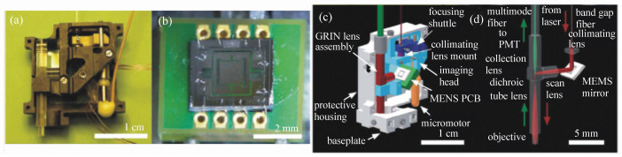
图 2 携带式双光子显微镜。(a)总览;(b)扫描镜整体焊线封装到印刷电路板上;(c)显微镜模型图(剖面观);(d)激光照明(红)和荧光收集(绿)光路[9]
Figure 2. Portable two-photon microscope. (a)overview; (b)the scanner die is wirebonded onto electrodes on the printed circuit board (PCB); (c)computer-aided-design model of the microscope, in a cut-away view; (d)laser illumination(red arrows) and fluorescence collection(green arrows) pathways[9]


表 1 近年报道的微型化双光子显微镜性能对比
Table 1. Comparison of miniaturized two-photon microscope′s characteristics reported recently
产品 头部佩戴重量 分辨率 扫描速度(频率、帧频) 扫描机制 Helmchen et al.(2001)[10] 25 g 亚细胞尺度 300~800 Hz(线扫描) 光纤探针扫描 Piyawattanametha et al.(2006)[12] - ~1 μm ~3.5 kHz(线扫描) MEMS扫描镜 Piyawattanametha et al.(2009)[9] 2.9 g 1.29±0.05 μm(横向)10.3±0.3 μm(轴向) 15 fps(光栅扫描)~1 kHz(线扫描) MEMS扫描镜 Zong et al. FHIRM-TPM(2017)[13] 2.15 g 0.64±0.02 μm(横向)3.35±0.37 μm(轴向) 40 fps(光栅扫描)10 kHz(线扫描) MEMS扫描镜  下载: 导出CSV
表 2 双光子与三光子成像部分特性的比较
Table 2. Comparison of selected features of two-photon and three-photon microscopy
下载: 导出CSV
-
[1] DENK W, STRICKLER J H, WEBB W W. Two-photon laser scanning fluorescence microscopy[J]. Science, 1990, 248(4951):73-76. doi: 10.1126/science.2321027 [2] DOMBECK D A, HARVEY C D, TIAN L, et al.. Functional imaging of hippocampal place cells at cellular resolution during virtual navigation[J]. Nature Neuroscience, 2010, 13(11):1433-1440. doi: 10.1038/nn.2648 [3] OLIVIER N, LUENGO-OROZ M A, DULOQUIN L, et al.. Cell lineage reconstruction of early zebrafish embryos using label-free nonlinear microscopy[J]. Science, 2010, 329(5994):967-971. doi: 10.1126/science.1189428 [4] MIZRAHI A, CROWLEY J C, SHTOYERMAN E, et al.. High-resolution in vivo imaging of hippocampal dendrites and spines[J]. Journal of Neuroscience, 2004, 24(13):3147-3151. doi: 10.1523/JNEUROSCI.5218-03.2004 [5] SVOBODA K, YASUDA R. Principles of two-photon excitation microscopy and its applications to neuroscience[J]. Neuron, 2006, 50(6):823-839. doi: 10.1016/j.neuron.2006.05.019 [6] NIELL C M, SMITH S J. Live optical imaging of nervous system development[J]. Annual Review of Physiology, 2004, 66(1):771-798. doi: 10.1146/annurev.physiol.66.082602.095217 [7] KERR J N, DENKW. Imaging in vivo:watching the brain in action[J]. Nature Reviews Neuroscience, 2008, 9(3):195-205. doi: 10.1038/nrn2338 [8] CHEN C C, LU J, ZUO Y. Spatiotemporal dynamics of dendritic spines in the living brain[J]. Frontires in Neuroanatomy, 2014, 8:28. http://cn.bing.com/academic/profile?id=bf3ed0a24a65f06fd1c6ae6d0b3214a0&encoded=0&v=paper_preview&mkt=zh-cn [9] PIYAWATTANAMETHA W, COCKER ED, BURNS L D, et al.. In vivo brain imaging using a portable 2.9 g two-photon microscope based on a microelectromechanical systems scanning mirror[J]. Optics Letters, 2009, 34(15):2309-2311. doi: 10.1364/OL.34.002309 [10] HELMCHEN F, FEE M S, TANK D W, et al.. A miniature head-mounted two-photon microscope:high-resolution brain imaging in freely moving animals[J]. Neuron, 2001, 31:903-912. doi: 10.1016/S0896-6273(01)00421-4 [11] MYAING M T, MACDONALD D J, LI X. Fiber-optic scanning two-photon fluorescence endoscope[J]. Optics Letters, 2006, 31(8):1076-1078. doi: 10.1364/OL.31.001076 [12] PIYAWATTANAMETHA W, BARRETTO R P J, KO T H, et al.. Fast-scanning two-photon fluorescence imaging based on a microelectromechanical systems two-dimensional scanning mirror[J]. Optics Letters, 2006, 31(13):2018-2020. doi: 10.1364/OL.31.002018 [13] ZONG W, WU R, LI M, et al.. Fast high-resolution miniature two-photon microscopy for brain imaging in freely behaving mice[J]. Nature Methods, 2017, 14(7):713-719. doi: 10.1038/nmeth.4305 [14] ATTARDO A, FITZGERALD J E, SCHNITZER M J. Impermanence of dendritic spines in live adult CA1 hippocampus[J]. Nature, 2015, 523(7562):592-596. doi: 10.1038/nature14467 [15] BOCARSLY M E, JIANG W C, WANG C, et al.. Minimally invasive microendoscopy system for in vivo functional imaging of deep nuclei in the mouse brain[J]. Biomedical Optics Express, 2015, 6(11):4546-4556. doi: 10.1364/BOE.6.004546 [16] CODA S, SIERSEMA P D, STAMP G W, et al.. Biophotonic endoscopy:a review of clinical research techniques for optical imaging and sensing of early gastrointestinal cancer[J]. Endoscopy International Open, 2015, 3(5):E380-392. doi: 10.1055/s-00025476 [17] GU M, BAO H, KANG H. Fibre-optical microendoscopy[J]. Journal of Microscopy, 2014, 254(1):13-18. doi: 10.1111/jmi.12119 [18] BIRD D, GU M. Fibre-optic two-photon scanning fluorescence microscopy[J]. Journal of Microscopy, 2002, 208:35-48. doi: 10.1046/j.1365-2818.2002.01059.x [19] BAO H C, ALLEN J, PATTIE R, et al.. Fast handheld two-photon fluorescence microendoscope with a 475μm×475μm field of view for in vivo imaging[J]. Optics Letters, 2008, 33(12):1333-1335. doi: 10.1364/OL.33.001333 [20] FU L, GU M. Fibre-optic nonlinear optical microscopy and endoscopy[J]. Journal of Microscopy, 2007, 226:195-206. doi: 10.1111/jmi.2007.226.issue-3 [21] FLUSBERG B A, COCKER E D, PIYAWATTANAMETHA W, et al.. Fiber-optic fluorescence imaging[J]. Nature Methods, 2005, 2(12):941-950. doi: 10.1038/nmeth820 [22] FLUSBERG B A, JUNG J C, COCKER E D, et al.. In vivo brain imaging using a portable 3.9 gram two-photon fluorescence microendoscope[J]. Optics Letters, 2005, 30(17):2272-2274. doi: 10.1364/OL.30.002272 [23] MYAING M T, YE J Y, NORRIS T B, et al.. Enhanced two-photon biosensing with double-clad photonic crystal fibers[J]. Optics Letters, 2003, 28(14):1224-1226. doi: 10.1364/OL.28.001224 [24] FU L, JAIN A, XIE H, et al.. Nonlinear optical endoscopy based on a double-clad photonic crystal fiber and a MEMS mirror[J]. Optics Express, 2006, 14(3):1027-1032. doi: 10.1364/OE.14.001027 [25] LIANG W X, HALL G, MESSERSCHMIDT B, et al.. Nonlinear optical endomicroscopy for label-free functional histology in vivo[J]. Light:Science & Applications, 2017, 6(7):e17082. https://www.researchgate.net/publication/320836063_Nonlinear_optical_endomicroscopy_for_label-free_functional_histology_in_vivo [26] AKINS M L, LUBY-PHELPS K, MAHENDROO M. Second harmonic generation imaging as a potential tool for staging pregnancy and predicting preterm birth[J]. Journal of Biomedical Optics, 2010, 15(2):026020. doi: 10.1117/1.3381184 [27] OHEIM M, BEAUREPAIRE E, CHAIGNEAU E, et al.. Two-photon microscopy in brain tissue:parameters influencing the imaging depth[J]. Journal of Neuroscience Methods, 2001, 111(1):29-37. doi: 10.1016/S0165-0270(01)00438-1 [28] CATALANO I M, CINGOLANI A. Three-photon absorption coefficient determination by means of nonlinear luminescence experiments[J]. Journal of Applied Physics, 1979, 50(9):5638-5641. doi: 10.1063/1.326738 [29] DAVEY A P, BOURDIN E, HENARI F, et al.. Three photon induced fluorescence from a conjugated organic polymer for infrared frequency upconversion[J]. Applied Physics Letters, 1995, 67(7):884-885. doi: 10.1063/1.114724 [30] HELL S F, BAHLMANN K, SCHRADER M, et al.. Three-photon excitation in fluorescence microscopy[J]. Journal of Biomedical Optics, 1996, 1(1):71-74. doi: 10.1117/12.229062 [31] GRYCZYNSKI I, SZMACINSKI H, LAKOWICZ J R. On the possibility of calcium imaging using indo-1 with three-photon excitation[J]. Photochemistry and Photobiology, 1995, 62(4):804-808. doi: 10.1111/php.1995.62.issue-4 [32] GRYCZYNSKI I, MALAK H, LAKOWICZ J R, et al.. Fluorescence spectral properties of troponin c mutant f22w with one-, two-, and three-photon excitation[J]. Biophysics Journal, 1996, 71(6):3448-3453. doi: 10.1016/S0006-3495(96)79540-1 [33] XU C, ZIPFEL W, SHEAR J B, et al.. Multiphoton fluorescence excitation:New spectral windows for biological nonlinear microscopy[J]. Proceedings of the National Academy of Sciences, 1996, 93:10763-10768. doi: 10.1073/pnas.93.20.10763 [34] WOKOSIN D L, CENTONZE V E, CRITTENDEN S, et al.. Three-photon excitation fluorescence imaging of biological specimens using an all-solid-state laser[J]. Bioimaging, 1996, 4:208-214. doi: 10.1002/1361-6374(199609)4:3<208::AID-BIO11>3.3.CO;2-A [35] MATSUDA H, FUJIMOTO Y, ITO S, et al.. Development of near-infrared 35 fs laser microscope and its application to the detection of three-and four-photon fluorescence of organic microcrystals[J]. The Journal of Physical Chemistry, 2006, 110:1091-1094. doi: 10.1021/jp0561165 [36] NORRIS G, AMOR R, DEMPSTER J, et al.. A promising new wavelength region for three-photon fluorescence microscopy of live cells[J]. Journal of Microscopy, 2012, 246(3):266-273. doi: 10.1111/j.1365-2818.2012.03610.x [37] HORTON N G, WANG K, KOBAT D, et al.. In vivo three-photon microscopy of subcortical structures within an intact mouse brain[J]. Nature Photonics, 2013, 7(3):205-209. doi: 10.1038/nphoton.2012.336 [38] ORON D, TAL E, SILBERBERG Y. Scanningless depth-resolved microscopy[J]. Optics Express, 2005, 13(5):1468-1476. doi: 10.1364/OPEX.13.001468 [39] ROWLANDS C J, PARK D, BRUNS O T, et al.. Wide-field three-photon excitation in biological samples[J]. Light:Science & Applications, 2016, 5:e16255. http://cn.bing.com/academic/profile?id=a8d6cfbf060c84ba943d8038832668ee&encoded=0&v=paper_preview&mkt=zh-cn [40] HELMCHEN F, DENK W. Deep tissue two-photon microscopy[J]. Nature Methods, 2005, 2(12):932-940. doi: 10.1038/nmeth818 [41] CHOI H, YEW E Y, HALLACOGLU B, et al.. Improvement of axial resolution and contrast in temporally focused widefield two-photon microscopy with structured light illumination[J]. Biomed Opt Express, 2013, 4(7):995-1005. doi: 10.1364/BOE.4.000995 -

 下载:
下载:


计量
- 文章访问数: 3352
- HTML全文浏览量: 1299
- PDF下载量: 455
- 被引次数: 0
