


-
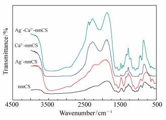
摘要: 为了提高壳聚糖的水溶性及其止血方面的性能,将壳聚糖(CS)纳米化,并引入具有抗菌作用的Ag+离子和凝血辅助作用的Ca2+离子,制备出纳米壳聚糖金属离子复合止血材料。首先,采用离子交换法制备纳米壳聚糖(nmCS),再分别加入AgNO3和饱和CaCl2溶液,制得nmCS-Ag、nmCS-Ca、nmCS-Ag-Ca复合材料。然后,采用FTIR、XRD、SEM等手段对复合材料的结构进行表征。最后,对复合物的凝血、止血性能进行了测试。实验结果表明:改性后的壳聚糖IR图谱在1 647 cm-1和1 560 cm-1处出现了纳米壳聚糖钠盐的特征吸收峰;复合了金属离子的纳米壳聚糖在XRD图谱中表现出了Ag+、Ca2+的晶型特征;扫描电镜显示nmCS-Ag中Ag+有部分析出而nmCS-Ca的复合效果较好;nmCS-Ag-Ca的凝血、止血效果要优于nmCS-Ag和nmCS-Ca,同时nmCS-Ag和nmCS-Ca的凝血、止血效果要优于nmCS。测试结果表明,成功制备了纳米壳聚糖金属离子复合止血材料。Abstract: In order to improve the water soluble and the hemostatic performance of chitosan (CS), chitosan is nano-sized and an antibacterial Ag+ and coagulation-assisted Ca2+ are introduced to prepare a nano-chitosan metal ion composite hemostatic material. Firstly, nano-chitosan (nmCS) is prepared by ion exchange method, and AgNO3 and saturated CaCl2 solution are added respectively to obtain nmCS-Ag, nmCS-Ca and nmCS-Ag-Ca composites. Then, the structure of the composite is characterized by means of FTIR, XRD and SEM. Finally, the coagulation and hemostatic properties of the complex are tested. The results show that the characteristic absorption peak of nano-chitosan sodium salt appeared at the modified chitosan IR spectrum at 1 647 cm-1 and 1 560 cm-1. The nano-chitosan composited with metal ions show the crystal characteristics of Ag+ and Ca2+ in the XRD pattern. The SEM shows that partial Ag+ precipitates in nmCS-Ag, and the composite effect of nmCS-Ca is better. The coagulation and hemostasis of nmCS-Ag-Ca is better than that of nmCS-Ag and nmCS-Ca, while the coagulation and hemostasis of nmCS-Ag and nmCS-Ca are better than that of nmCS. The test results show that the nano-chitosan metal ion composite hemostatic material has been successfully prepared.
-
Key words:
- nano-chitosan /
- composite materials /
- characterization /
- performance
-

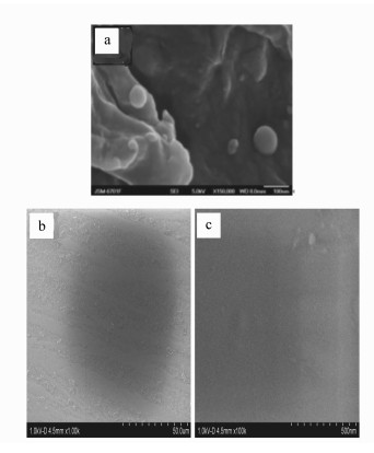
图 5 nmCS、Ca-nmCS及Ag-nmCS扫描电镜图
Figure 5. SEM micrographs of nm Chitosan composite Ag+、Ca2+
表 1 样品的凝血时间测试
Table 1. Clotting time test of the sample
样品 编号 凝血时间 平均凝血时间 自然凝血 1 5′37″ 5′18″ 2 5′2″ 3 5′16″ CS 1 3′40″ 3′56″ 2 3′23″ 3 3′40″ nmCS 1 3′23″ 3′27″ 2 3′20″ 3 3′37″ Ag+-nmCS 1 3′7″ 3′17″ 2 3′25″ 3 3′19″ Ca2+-nmCS 1 3′22″ 3′13″ 2 3′14″ 3 3′3″ Ag+-Ca2+-nmCS 1 2′55″ 3′2″ 2 3′7″ 3 3′5″  下载: 导出CSV
表 2 样品的止血时间测试
Table 2. Bleeding time test of the sample
样品 编号 止血时间 平均止血时间 自然止血 1 18′37″ 17′34″ 2 16′24″ 3 17′40″ CS 1 9′25″ 9′46″ 2 10′5″ 3 9′48″ 9′24″ nmCS 1 9′40″ 2 9′8″ 3 9′25″ Ag+- nmCS 1 8′40″ 8′37″ 2 8′23″ 3 8′49″ Ca2+-nmCS 1 8′28″ 8′9″ 2 7′45″ 3 8′13″ Ag+-Ca2+-nmCS 1 7′36″ 7′12″ 2 6′46″ 3 7′15″
下载: 导出CSV
-
[1] PANWAR P, LAMOUR G, MACKENZIE N C W, et al..Changes in structural-mechanical properties and degradability of collagen during aging-associated modifications[J]. Journal of Biological Chemistry, 2015, 290(38):23291-23306. doi: 10.1074/jbc.M115.644310 [2] SALGADO C L, GRENHO L, FERNANDES M H, et al.. Biodegradation, biocompatibility, and osteoconduction evaluation of collagen Nanohydroxyapatite cryogels for bone tissue regeneration[J]. Journal of Biomedical Materials Research Part A, 2016, 104(1):57-70. doi: 10.1002/jbm.a.35540 [3] GU RL, SUN WZ, ZHOU H, et al.. The performance of a fly-larva shell-derived chitosan sponge as an absorbable surgical hemostatic agent[J]. Biomaterials, 2010, 31(6):1270-1277. doi: 10.1016/j.biomaterials.2009.10.023 [4] 李学军, 孙园园.不同生物止血材料研究进展及复合型止血材料的临床应用[J].中国组织工程研究与临床康复, 2011, 15(51):9671-9674. doi: 10.3969/j.issn.1673-8225.2011.51.041LI X J, SUN Y Y. Advances in various biomaterials for hemostasis and clinical application of compound materials for hemostasis[J]. Journal of Clinical Rehabilitative Tissue Engineering Research, 2011, 15(51):9671-9674.(in Chinese) doi: 10.3969/j.issn.1673-8225.2011.51.041 [5] 胡帼颖, 顾汉卿.止血剂基础与临床应用研究新进展[J].透析与人工器官, 2010, 21(2):29-35. http://d.old.wanfangdata.com.cn/Periodical/txyrgqg201002007HU G Y, GU H Q. New research on the basis and clinical application of hemostatic agents[J]. Chinese J. Dial. & Artif. Organs, 2010, 21(2):29-35.(in Chinese) http://d.old.wanfangdata.com.cn/Periodical/txyrgqg201002007 [6] ONG S Y, WU J, MOOCHHALA S M, et al..Development of achitosan-based wound dressing with improved hemostatic and antimicrobial properties[J]. Biomaterials, 2008, 29(32):4323-4332. doi: 10.1016/j.biomaterials.2008.07.034 [7] 杜建华, 刘彦伟, 李园园.纳米颗粒增强酮基摩擦材料的摩擦学性能[J].光学精密工程, 2013, 21(10):2581-2586. http://kns.cnki.net/KCMS/detail/detail.aspx?filename=GXJM201310016&dbname=CJFD&dbcode=CJFQDU J H, LIU Y W, LI Y Y. Nanoparticles enhance the tribological properties of ketone-based friction materials[J]. Opt. Precision Eng., 2013, 21(10):2581-2586.(in Chinese) http://kns.cnki.net/KCMS/detail/detail.aspx?filename=GXJM201310016&dbname=CJFD&dbcode=CJFQ [8] 孙磊.高粘度壳聚糖止血材料的制备及其应用性研究[D].北京: 中央民族大学, 2012. http://cdmd.cnki.com.cn/Article/CDMD-10052-1012416220.htmSUN L. Preparation and application of high viscosity chitosan hemostatic material[D]. Beijing: Minzu University of China, 2012.(in Chinese) http://cdmd.cnki.com.cn/Article/CDMD-10052-1012416220.htm [9] STEFANI I, COOPER-WHITE J J. Development of an in-process UV-crosslinked, electrospun PCL/a PLA-co-TMC composite polymer for tubular tissue engineering applications[J]. Acta Biomaterialia, 2016, 36:231-240. doi: 10.1016/j.actbio.2016.03.013 [10] 丁萍, 黄可龙, 刘艳飞, 等.壳聚糖衍生物与Ca(Ⅱ)螯合反应的动力学及机理研究[J].动能材料, 2008, 39(6):961-964. http://www.wanfangdata.com.cn/details/detail.do?_type=perio&id=gncl200806024DING P, HUANG K L, LIU Y F, et al.. Kinetics and mechanism research of chitosan derivatives with Ca(Ⅱ) chelate reaction[J]. Kinetic Energy Materials, 2008, 39(6):961-964. http://www.wanfangdata.com.cn/details/detail.do?_type=perio&id=gncl200806024 [11] ZHOU Z L, SHANG H B, SUI Y X, et al.. Useful way to compensate for intrinsic birefringence caused by calcium fluoride in optical lithography systems[J]. Chinese Optic Letters, 2018, 16(3), 032201:1-5. http://www.cnki.com.cn/Article/CJFDTotal-GXKB201803016.htm [12] RODRIGUEZ N R M, DAS S, KAUFMAN Y, et al.. Mussel adhesive protein provides cohesive matrix for collagen type-1α[J]. Biomaterials, 2015, 51:51-57. doi: 10.1016/j.biomaterials.2015.01.033 [13] FANG J Q, JIANG L, CAO Q, et al.. Doping transition metal ions as a method for enhancement of ablation rate in femtosecond laser irradiation of silicate glass[J]. Chinese Optic Letters, 2014, 12, 120402:1-5. http://www.opticsinfobase.org/col/abstract.cfm?uri=col-12-12-121402 [14] 许辉, 张俊举, 袁轶慧, 等.红外与可见光图像融合系统的探测概率[J].光学精密工程, 2013, 21(12):3205-3213. http://d.old.wanfangdata.com.cn/Periodical/gxjmgc201312030XU H, ZHANG J J, YUAN Y H, et al.. Detection probability of infrared and visible image fusion system[J]. Opt. Precision Eng., 2013, 21(12):3205-3213.(in Chinese) http://d.old.wanfangdata.com.cn/Periodical/gxjmgc201312030 -

 下载:
下载:


图(5) / 表(2)
计量
- 文章访问数: 3103
- HTML全文浏览量: 1462
- PDF下载量: 174
- 被引次数: 0
