


-
摘要: 皮肤镜在临床中已广泛用于诊断皮肤病变,然而,如何更加清晰准确地观察表皮、真皮表皮交界处和真皮乳头层内的色素性结构的大小、形状、颜色的深浅及浅层血管丛血管的大小形态依旧是一个挑战。由于皮肤表皮层类似于镜面反射,不改变反射光的偏振态,而皮肤组织表皮层以下的组织会改变原有光线的偏振态,基于此,本文设计了一个视场为22 mm、倍率为10×的手持偏振皮肤镜,通过采用偏振照明成像技术可以更加清晰有效地观察深层皮肤组织的病变以及病变组织形态,从而提高临床诊断与鉴别效率。Abstract: Dermoscopy has been widely used in the diagnosis of skin lesions in the clinical field. However, difficulty remains in clear and accurate observation of the size, shape, color depth and size of superficial vascular plexus vessels in the epidermis, dermal-epidermal junction and dermal papilla. Since the epidermis is specularly reflective, it does not change the polarization state of the reflected light. Contrarily, tissue below the epidermal layer of the skin tissue changes the polarization state of the source light. Based on this, we designed a handheld system with a field of view of 22 mm and a magnification of 10×that performs polarized dermoscopy through the use of polarized illumination imaging technology, which more clearly and effectively observe the lesions of deep skin tissue and the morphology of lesions, thereby improving the efficiency of clinical diagnoses.
-
Key words:
- dermoscopy /
- skin disease imagery /
- polarized light
-
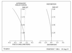
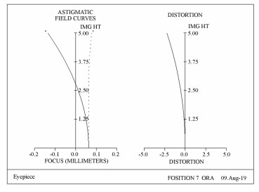
图 5 偏振皮肤镜场曲和畸变曲线
Figure 5. Field curvature and distortion curve of polarized dermoscopy
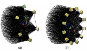
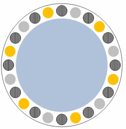
图 7 LightTools模拟光源照度均匀性。(a)模拟6个白色LED光源照度;(b)模拟12个白色LED偏振光源照度
Figure 7. Illuminance uniformity of the source simulated by LightTools. (a) Simulate illumination of 6 white LED sources; (b) simulate illumination of 12 white LEDs with polarized light source
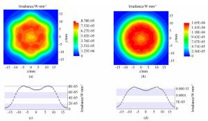
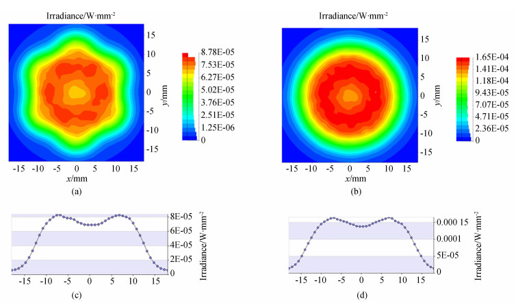
图 8 非偏振与偏振LED光源照度图。(a)非偏振光源照度图;(b)偏振光源照度图;(c)非偏振光源Y轴辐射分布;(d)偏振光源Y轴辐射分布
Figure 8. Unpolarized and polarized LED source illuminations. (a) Unpolarized source illumination; (b) polarized source illumination; (c) Y-axis radiation distribution of unpolarized source; (d) Y-axis radiation distribution of polarized source.
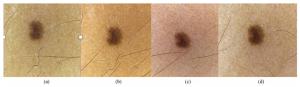
图 10 (a) 白光非偏振下的皮损形态;(b)黄光非偏振下的皮损形态;(c)白光偏振下的皮损形态;(d)黄光偏振下的皮损形态
Figure 10. (a) Skin lesion under white unpolarized light; (b) skin lesion under yellow unpolarized light; (c) skin lesion under white polarized light; (d) skin lesion under yellow polarized light.
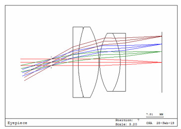
表 1 最终目镜结构参数
Table 1. Structure parameters of final eyepiece
参数名称 参数 口径/mm 30 光学总长/mm 28.81 焦距/mm 25 入瞳直径/mm 4 目视倍率 10× 有效靶面/mm 22  下载: 导出CSV
表 2 模拟LED光源照明参数
Table 2. Simulation LED lighting parameters
设置条件 白色LED光源参数 偏振LED光源参数 LED数目 6 12 LED发散角 30° 30° LED倾斜角度 25° 25° 对角LED距离 35 mm 35 mm LED距靶面距离 19 mm 19 mm 照明均匀性 85% 86%
下载: 导出CSV
-
[1] Cancer facts & figures 2009[EB/OL]. The American Cancer Society.[2010-08-18]. http://www.cancer.org. [2] ZALAUDEK I, ARGENZIANO G, DI STEFANI A, et al.. Dermoscopy in general dermatology[J]. Dermatology, 2006, 212(1):7-18. http://cn.bing.com/academic/profile?id=67b2569d85603ceb0d4d0fbc8cbd7a49&encoded=0&v=paper_preview&mkt=zh-cn [3] BRAUN R P, RABINOVITZ H S, OLIVIERO M, et al.. Dermoscopy of pigmented skin lesions[J]. Journal of the American Academy of Dermatology, 2005, 52(1):109-121. doi: 10.1016/j.jaad.2001.11.001 [4] MENZIES S W, CROTTY K A, C INGVAR C, et al.. Dermoscopy:An Atlas[M]. 3rd ed. Sydney, Australia:McGraw-Hill Book Co, 2009:26-32. [5] MARGHOOB A A, BRAUN R P, KOPF A W. Atlas of Dermoscopy[M]. London:Taylor & Francis, 2005:374. [6] MALVEHY J, PUIG S, BRAUN R P, et al.. Handbook of Dermoscopy[M]. London:Taylor & Francis, 2006:88-90. [7] YAROSLAVSKY A N, NEEL V, ANDERSON R R. Fluorescence polarization imaging for delineating nonmelanoma skin cancers[J]. Optics Letters, 2004, 29(17):2010-2012. doi: 10.1364/OL.29.002010 [8] 孟如松, 蔡瑞康, 赵广, 等.偏振光皮肤镜数字图像分析技术的研究及在色素性皮损诊断中的探讨[J].中国体视学与图像分析, 2006, 11(2):122-126. doi: 10.3969/j.issn.1007-1482.2006.02.014MENG R S, CAI R K, ZHAO G, et al.. Study on digital image analysis technique of polarized dermabrasion and its application in the diagnosis of pigmented lesions[J]. Chinese Journal of Stereology and Image Analysis, 2006, 11(2):122-126. (in Chinese) doi: 10.3969/j.issn.1007-1482.2006.02.014 [9] BENVENUTO-ANDRADE C, DUSZA S W, AGERO A L C, et al.. Differences between polarized light dermoscopy and immersion contact dermoscopy for the evaluation of skin lesions[J]. Archives of Dermatology, 2007, 143(3):329-338. http://www.wanfangdata.com.cn/details/detail.do?_type=perio&id=2742f1d0d64b917e48a39f60300052eb [10] 马科斯·波恩, 埃米尔·沃耳夫.光学原理[M].杨葭荪, 译.北京: 电子工业出版社, 2005: 34-36.BONN M, WOLF E. Principles of Optics[M]. YANG X S, trans. Beijing: Publishing House of Electronics Industry, 2005: 34-36. (in Chinese) [11] 吴亚飞, 顾洪恩, 李增智, 等.大学物理(下册)[M].北京:高等教育出版社, 2015:140.WU Y F, GU H E, LI Z ZH, et al.. University Physics (2)[M]. Beijing:Higher Education Press, 2015:140. (in Chinese) [12] ANDERSON R R, PARRISH J A. The optics of human skin[J]. Journal of Investigative Dermatology, 1981, 77(1):13-19. doi: 10.1111-1523-1747.ep12479191/ [13] ZONIOS G, BYKOWSKI J, KOLLIAS N. Skin melanin, hemoglobin, and light scattering properties can be quantitatively assessed in vivo using diffuse reflectance spectroscopy[J]. Journal of Investigative Dermatology, 2001, 117(6):1452-1457. doi: 10.1046/j.0022-202x.2001.01577.x [14] TSENG S H, BARGO P, DURKIN A, et al.. Chromophore concentrations, absorption and scattering properties of human skin in-vivo[J]. Optics Express, 2009, 17(17):14599-14617. doi: 10.1364/OE.17.014599 -

 下载:
下载:


计量
- 文章访问数: 2556
- HTML全文浏览量: 1824
- PDF下载量: 165
- 被引次数: 0
