Improving sensitivity by multi-coherence of magnetic surface plasmons
doi: 10.37188/CO.EN.2022-0009
-
摘要:
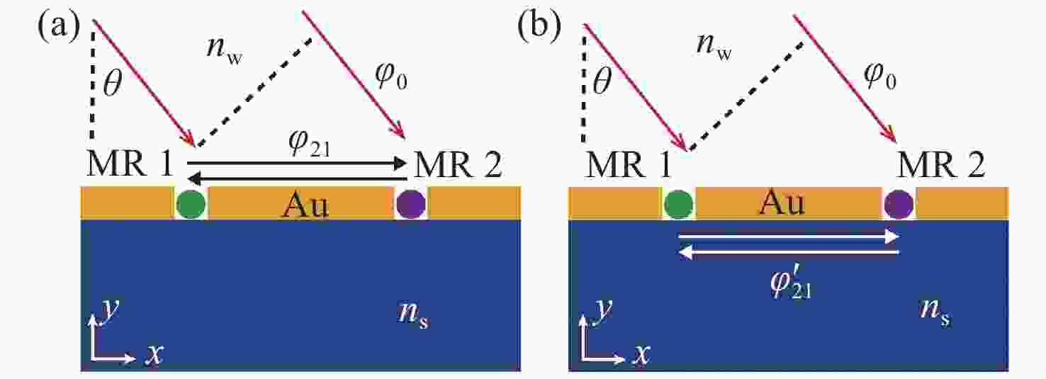
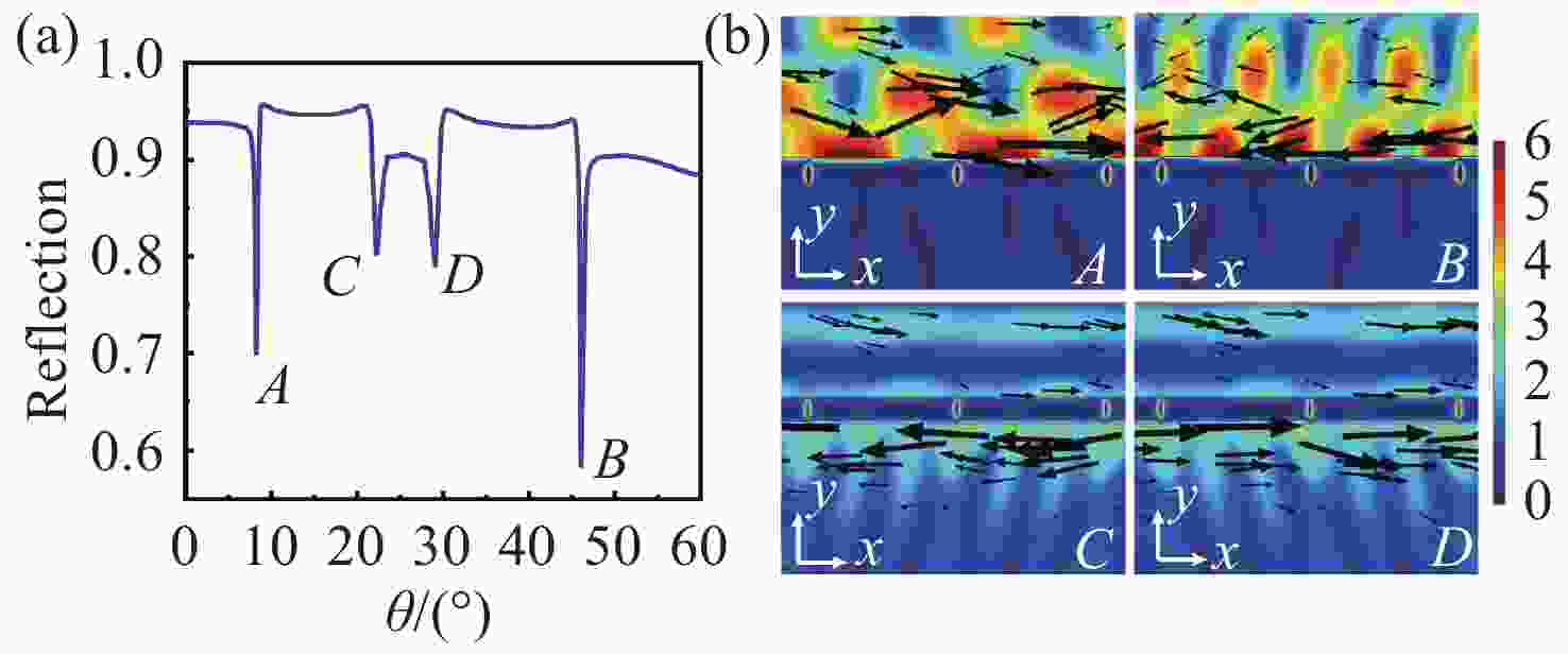
本文研究了一维金属纳米狭缝阵列中磁表面等离激元的相干现象,并提出了一种双谷传感方法以提高灵敏度。与通常所采用的固定入射角度进行波长扫描的方式不同,本文采用固定波长改变入射角度的方式研究表面等离激元的相干现象。由于延迟效应的存在,随着周围介质折射率的变化,两个谷会向相反的方向移动。相比于使用单一谷进行标定的方式,两个相反方向移动的谷可以有效提高灵敏度。用于标定的两个谷单独的灵敏度最大分别为39.2°/RIU和102.4°/RIU,而双谷标定的总灵敏度可达141.6°/RIU。此外,狭缝介质与上层介质的折射率不一致对传感性能的影响很小,故其有广泛的应用前景。
Abstract:In this paper, we study the coherence of magnetic surface plasmons in one-dimensional metallic nano-slit arrays and propose a double-dip sensing method to improve sensitivity. Different from the conventional way of scanning wavelength at a fixed incident angle, coherence of surface plasmons is investigated by changing the incident angle at a fixed wavelength. Due to the retardation effect, two coherence dips move in opposite directions as the refractive index of the surrounding medium changes. Compared with one dip used for sensing, two oppositely moving dips can efficiently improve the sensitivity. The total sensitivity of two dips can reach 141.6°/RIU while the sensitivities of two single dips are 39.2°/RIU and 102.4°/RIU respectively. Besides, the inconsistency between the refractive index of slit medium and upper medium has few influences on the sensing performance, which will have wide practical applications.
-
Key words:
- surface plasmons /
- refractive index sensor /
- coherence /
- metal grating
-
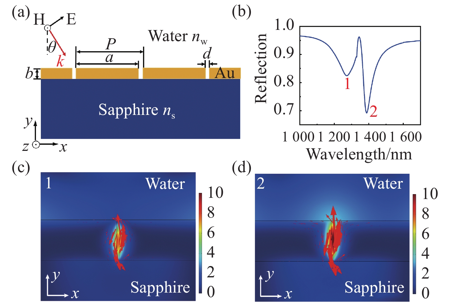
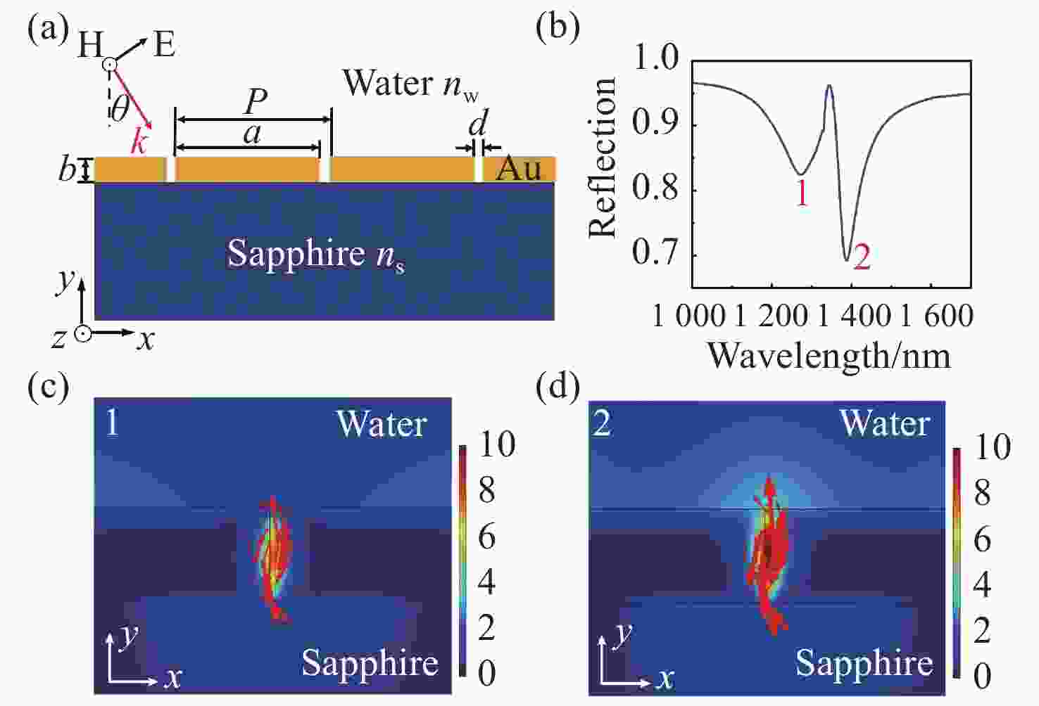
Figure 1. (a) Schematic diagram of one-dimensional metallic nano-slit arrays structure on sapphire substrate. The whole structure is immersed in water; (b) the reflection spectrum at normal incidence; (c)(d) the normalized magnetic field intensity distributions corresponding to the dip 1 and dip 2, respectively
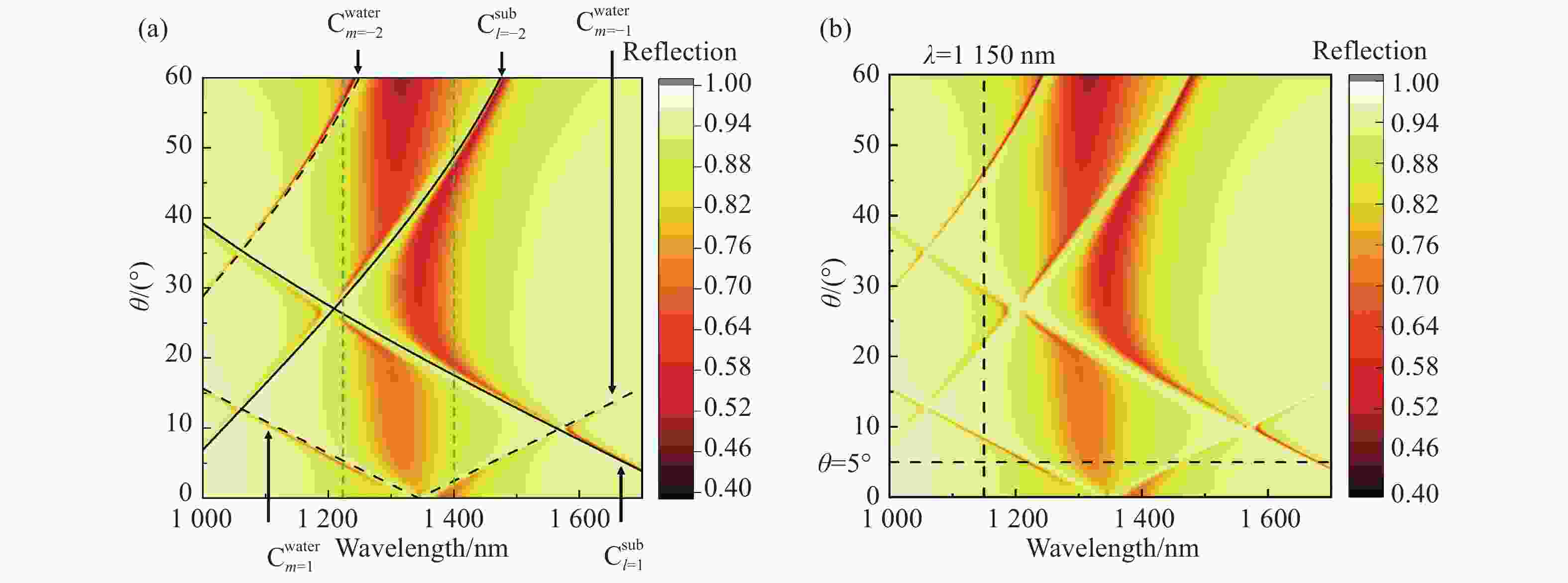
Figure 3. 2D Reflection spectra of the structure in the range of
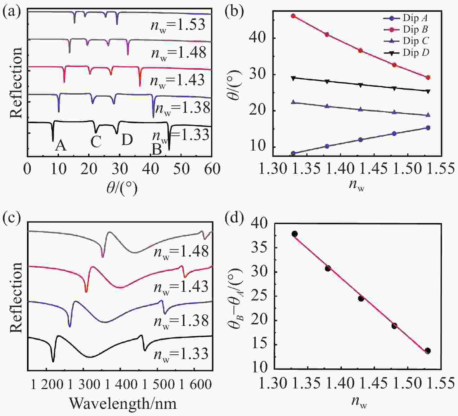
$\theta = 0 \sim 60^\circ $ and$\lambda = 1\;000 \sim 1\;700\;{\rm{nm}}$ . (a) Three dashed lines$C_{m = - 1}^{{\rm{water}}}$ ,$C_{m = 1}^{{\rm{water}}}$ , and$C_{m = - 2}^{{\rm{water}}}$ represent different orders of magnetic surface plasmon coherence corresponding to the interface between metal and water, respectively. Two solid lines$C_{l = - 2}^{{\rm{sub}}}$ and$C_{l = 1}^{{\rm{sub}}}$ represent different orders of coherence corresponding to the interface between metal and substrate; (b) positions of fixed wavelength$\lambda = 1150\;{\rm{nm}}$ and fixed incident angle$\theta = 5^\circ $ are marked by vertical and horizontal dashed lines in 2D spectrum, respectivelyFigure 5. (a) The angle-resolved reflectance spectrum obtained by changing the refractive index of the upper medium (nw=1.33 ~ 1.53) with an interval of 0.05 at the wavelength of 1150 nm. (b) Dip position varying with the refractive index. (c) Reflection spectrum is obtained by changing the wavelength when
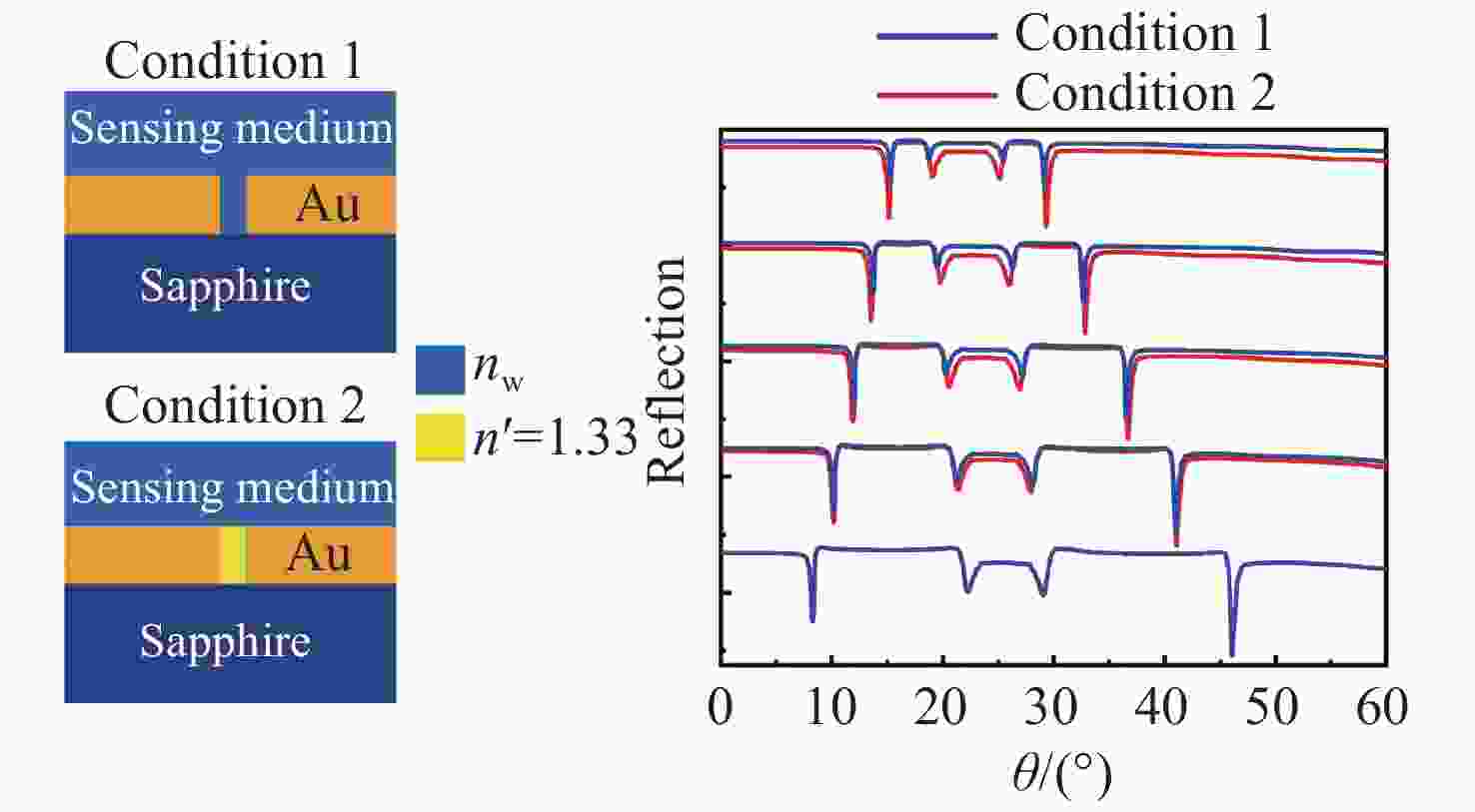
$\theta $ is fixed at 5°. As the refractive index increases, both coherence dips are red-shifted. (d) The angle difference between dip A and dip B varying with the upper mediumTable 1. Sensitivities and FOMs with different refractive indices
n 1.33 1.38 1.43 1.48 1.53 SA/(°)·RIU−1 39.2 37.4 34.8 33.2 32.4 SB/(°)·RIU−1 102.4 95.4 83.4 74.2 70 S/(°)·RIU−1 141.6 132.8 118.2 107.4 102.4 FOMA/RIU−1 115.2 104.0 91.8 85.6 82.9 FOMB/RIU−1 214.1 213.7 185.9 172.3 167.3 -
[1] KOYA A N, ZHU X CH, OHANNESIAN N, et al. Nanoporous metals: from plasmonic properties to applications in enhanced spectroscopy and photocatalysis[J]. ACS Nano, 2021, 15(4): 6038-6060. doi: 10.1021/acsnano.0c10945 [2] HE ZH H, XUE W W, CUI W, et al. Tunable fano resonance and enhanced sensing in a simple Au/TiO2 hybrid metasurface[J]. Nanomaterials, 2020, 10(4): 687. doi: 10.3390/nano10040687 [3] PALERMO G, SREEKANTH K V, MACCAFERRI N, et al. Hyperbolic dispersion metasurfaces for molecular biosensing[J]. Nanophotonics, 2021, 10(1): 295-314. [4] AHMADIVAND A, GERISLIOGLU B, RAMEZANI Z, et al. Functionalized terahertz plasmonic metasensors: femtomolar-level detection of SARS-CoV-2 spike proteins[J]. Biosensors and Bioelectronics, 2021, 177: 112971. doi: 10.1016/j.bios.2021.112971 [5] SHEN B L, LIU L W, LI Y P, et al. Nonlinear spectral-imaging study of second- and third-harmonic enhancements by surface-lattice resonances[J]. Advanced Optical Materials, 2020, 8(8): 1901981. doi: 10.1002/adom.201901981 [6] GUAN J, SAGAR L K, LI R, et al. Quantum dot-plasmon lasing with controlled polarization patterns[J]. ACS Nano, 2020, 14(3): 3426-3433. doi: 10.1021/acsnano.9b09466 [7] DORRAH A H, CAPASSO F. Tunable structured light with flat optics[J]. Science, 2022, 376(6591): eabi6860. doi: 10.1126/science.abi6860 [8] PANDEY P S, RAGHUWANSHI S K, KUMAR S. Recent advances in two-dimensional materials-based kretschmann configuration for SPR sensors: a review[J]. IEEE Sensors Journal, 2022, 22(2): 1069-1080. doi: 10.1109/JSEN.2021.3133007 [9] XUE T Y, LIANG W Y, LI Y W, et al. Ultrasensitive detection of miRNA with an antimonene-based surface plasmon resonance sensor[J]. Nature Communications, 2019, 10(1): 28. doi: 10.1038/s41467-018-07947-8 [10] BAGHBADORANI H K, BARVESTANI J. Sensing improvement of 1D photonic crystal sensors by hybridization of defect and Bloch surface modes[J]. Applied Surface Science, 2021, 537: 147730. doi: 10.1016/j.apsusc.2020.147730 [11] LIU F X, SONG B X, SU G X, et al. Sculpting extreme electromagnetic field enhancement in free space for molecule sensing[J]. Small, 2018, 14(33): 1801146. doi: 10.1002/smll.201801146 [12] CATTONI A, GHENUCHE P, HAGHIRI-GOSNET A M, et al. λ3/1000 plasmonic nanocavities for biosensing fabricated by soft UV nanoimprint lithography[J]. Nano Letters, 2011, 11(9): 3557-3563. doi: 10.1021/nl201004c [13] MAYER K M, HAFNER J H. Localized surface plasmon resonance sensors[J]. Chemical Reviews, 2011, 111(6): 3828-3857. doi: 10.1021/cr100313v [14] NUGROHO F A A, ALBINSSON D, ANTOSIEWICZ T J, et al. Plasmonic metasurface for spatially resolved optical sensing in three dimensions[J]. ACS Nano, 2020, 14(2): 2345-2353. doi: 10.1021/acsnano.9b09508 [15] BUKASOV R, SHUMAKER-PARRY J S. Highly tunable infrared extinction properties of gold nanocrescents[J]. Nano Letters, 2007, 7(5): 1113-1118. doi: 10.1021/nl062317o [16] HOU Y M. Coherence of magnetic resonators in a metamaterial[J]. AIP Advances, 2013, 3(12): 122119. doi: 10.1063/1.4861028 [17] HOU Y M. Interaction of magnetic resonators studied by the magnetic field enhancement[J]. AIP Advances, 2013, 3(12): 122118. doi: 10.1063/1.4861027 [18] KRAVETS V G, SCHEDIN F, GRIGORENKO A N. Extremely narrow plasmon resonances based on diffraction coupling of localized plasmons in arrays of metallic nanoparticles[J]. Physical Review Letters, 2008, 101(8): 087403. doi: 10.1103/PhysRevLett.101.087403 [19] LIMONOV M F, RYBIN M V, PODDUBNY A N, et al. Fano resonances in photonics[J]. Nature Photonics, 2017, 11(9): 543-554. doi: 10.1038/nphoton.2017.142 [20] LIMONOV M F. Fano resonance for applications[J]. Advances in Optics and Photonics, 2021, 13(3): 703-771. doi: 10.1364/AOP.420731 [21] XIAO SH Y, WANG T, LIU T T, et al. Active metamaterials and metadevices: a review[J]. Journal of Physics D:Applied Physics, 2020, 53(50): 503002. doi: 10.1088/1361-6463/abaced [22] WANG B Q, YU P, WANG W H, et al. High-Q plasmonic resonances: fundamentals and applications[J]. Advanced Optical Materials, 2021, 9(7): 2001520. doi: 10.1002/adom.202001520 [23] UTYUSHEV A D, ZAKOMIRNYI V I, RASSKAZOV I L. Collective lattice resonances: plasmonics and beyond[J]. Reviews in Physics, 2021, 6: 100051. doi: 10.1016/j.revip.2021.100051 [24] KRAVETS V G, KABASHIN A V, BARNES W L, et al. Plasmonic surface lattice resonances: a review of properties and applications[J]. Chemical Reviews, 2018, 118(12): 5912-5951. doi: 10.1021/acs.chemrev.8b00243 [25] DONG J W, CHEN SH, HUANG G F, et al. Low-index-contrast dielectric lattices on metal for refractometric sensing[J]. Advanced Optical Materials, 2020, 8(21): 2000877. doi: 10.1002/adom.202000877 [26] CHEN J, ZHANG Q, PENG CH, et al. Optical cavity-enhanced localized surface plasmon resonance for high-quality sensing[J]. IEEE Photonics Technology Letters, 2018, 30(8): 728-731. doi: 10.1109/LPT.2018.2814216 [27] LIANG L, ZHENG Q L, WEN L, et al. Miniaturized spectroscopy with tunable and sensitive plasmonic structures[J]. Optics Letters, 2021, 46(17): 4264-4267. doi: 10.1364/OL.426624 [28] WANG H, WANG X L, YAN CH, et al. Full color generation using silver tandem nanodisks[J]. ACS Nano, 2017, 11(5): 4419-4427. doi: 10.1021/acsnano.6b08465 [29] DAQIQEH REZAEI S, DONG ZH G, YOU EN CHAN J, et al. Nanophotonic structural colors[J]. ACS Photonics, 2021, 8(1): 18-33. doi: 10.1021/acsphotonics.0c00947 [30] SHI X ZH, CHEN CH SH, LIU S H, et al. Nonvolatile, reconfigurable and narrowband mid-infrared filter based on surface lattice resonance in phase-change Ge2Sb2Te5[J]. Nanomaterials, 2020, 10(12): 2530. doi: 10.3390/nano10122530 [31] MURAVITSKAYA A, GOKARNA A, MOVSESYAN A, et al. Refractive index mediated plasmon hybridization in an array of aluminium nanoparticles[J]. Nanoscale, 2020, 12(11): 6394-6402. doi: 10.1039/C9NR09393A [32] SHEN Y, ZHOU J H, LIU T R, et al. Plasmonic gold mushroom arrays with refractive index sensing figures of merit approaching the theoretical limit[J]. Nature Communications, 2013, 4(1): 2381. doi: 10.1038/ncomms3381 [33] LINDEN S, ENKRICH C, WEGENER M, et al. Magnetic response of metamaterials at 100 terahertz[J]. Science, 2004, 306(5700): 1351-1353. doi: 10.1126/science.1105371 [34] ZHU Y H, ZHANG H, LI D M, et al. Magnetic plasmons in a simple metallic nanogroove array for refractive index sensing[J]. Optics Express, 2018, 26(7): 9148-9154. doi: 10.1364/OE.26.009148 [35] CHEN J, FAN W F, ZHANG T, et al. Engineering the magnetic plasmon resonances of metamaterials for high-quality sensing[J]. Optics Express, 2017, 25(4): 3675-3681. doi: 10.1364/OE.25.003675 [36] CHEN X SH, PARK H R, LINDQUIST N C, et al. Squeezing millimeter waves through a single, nanometer-wide, centimeter-long slit[J]. Scientific Reports, 2014, 4(1): 6722. [37] RHIE J, LEE D, BAHK Y M, et al. Control of optical nanometer gap shapes made via standard lithography using atomic layer deposition[J]. Journal of Micro/Nanolithography,MEMS,and MOEMS, 2018, 17(2): 023504. [38] JOHNSON P B, CHRISTY R W. Optical constants of the noble metals[J]. Physical Review B, 1972, 6(12): 4370-4379. doi: 10.1103/PhysRevB.6.4370 [39] HOSSAIN M B, RANA M M, ABDULRAZAK L F, et al. Design and analysis of graphene–MoS2 hybrid layer based SPR biosensor with TiO2–SiO2 nano film for formalin detection: numerical approach[J]. Optical and Quantum Electronics, 2019, 51(6): 195. doi: 10.1007/s11082-019-1911-z [40] PANDEY P S, SINGH Y, RAGHUWANSHI S K. Theoretical analysis of the LRSPR sensor with enhance FOM for low refractive index detection using MXene and fluorinated graphene[J]. IEEE Sensors Journal, 2021, 21(21): 23979-23986. doi: 10.1109/JSEN.2021.3112530 [41] MUDGAL N, SAHARIA A, AGARWAL A, et al. ZnO and Bi-metallic (Ag–Au) layers based surface plasmon resonance (SPR) biosensor with BaTiO3 and graphene for biosensing applications[J]. IETE Journal of Research, 2020: 1-8. doi: 10.1080/03772063.2020.1844074 [42] HOSSAIN M M, TALUKDER M A. Gate-controlled graphene surface plasmon resonance glucose sensor[J]. Optics Communications, 2021, 493: 126994. doi: 10.1016/j.optcom.2021.126994 -


 下载:
下载:
